| Title |
Traité d'anatomie pathologique générale et spéciale : ou description et iconographie pathologique des altérations morbides tant liquides que solides observées dans le corps humain: Atlas, volume 1 |
| Call Number |
RB24 .L42; Record ID 99136490102001 |
| Date |
1857 |
| Description |
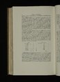
Hermann Lebert studied medicine in Berlin, Zürich, and Paris, where he also practiced medicine. He was one of the first anatomists of the nineteenth century to use the microscope for pathological diagnostic studies. In his research, he distinguished between tuberculosis and cancer. In Traité D'Anatomie Pathologique, Générale et Spéciale, Lebert was one of the first to describe premalignant polyps of the colon, rectum, and stomach. Traité D'Anatomie Pathologique was the most comprehensive illustrated medical dictionary to date, containing clinical material from the best-regarded physicians of France. The illustrations are steel-face copper engravings printed in color using multiple runs by the most renowned publishing house of the time. |
| Creator |
Lebert, Hermann, 1813-1878 |
| Subject |
Anatomy, Pathological |
| Type |
Text |
| Format |
application/pdf |
| Language |
fra |
| Collection Name |
Rare Books Collection |
| Holding Institution |
Rare Books Division, Special Collections, J. Willard Marriott Library, University of Utah |
| Rights |
 |
| Scanning Technician |
Natalia Soto |
| ARK |
ark:/87278/s6769wd9 |
| Setname |
uum_rbc |
| ID |
1424042 |
| Reference URL |
https://collections.lib.utah.edu/ark:/87278/s6769wd9 |