OCR Text |
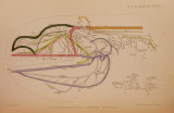
Show NERVES OF CHLAMYDOSELACHUS ANGUINEUS. 9 8 3 from the pre-trematic in the usual manner. The post-trematic passes along the sixth arch, sending out no branches, but unites, halfway along the arch, with a fine nerve (PI. LXVIII. fig. 1, V. 6), probably Vagus 6. This combined nerve (fig. 1, Y. 5 + V. 6) could be traced along the arch as far as the middle of the cerato-branchial cartilage, where it divides into a number of small branches which supply the arch and muscles attached to it. No fibres have been traced on to the vestigial seventh arch. On one side, not only do post-trematic 5 and Vagus 6 unite completely, but they are also connected by a transverse branch (fig. 1, Con. V. 5). (</) When the nerve called Vagits 6 is traced upward, it is found to originate from the vagal riband, between the origin of Vagus 5 and the point where the intestinal trunk divides. Traced from its central connections Vagus 6 passes obliquely downward and caudad, to fork over the anterior cardinal. The anterior part unites with the post-trematic of Vagus 5 and the posterior with one of the first spinal nerves. The origin, course, and distribution of this nerve strongly suggests that it is the remnant of the sixth branchial nerve to the degenerate seventh branchial arch. It is highly probable that serial sections would show that the combined nerve sends branches not only to the sixth arch but also to the vestigial seventh arch, which lies in close proximity to the former. The posterior division of Vagus 6 is very small and unites with a spinal nerve (or nerves), whose main distribution is to the median mandibular muscles. The anterior cardinal vein in Chlamydoselachus lies in the position of the vanished seventh cleft, but in most Elasmobranchs in the position of the missing sixth cleft. In the latter, the vein sometimes lies in the notch, so common on the fifth branchial arch, which in many cases is undoubtedly produced by a remnant of the sixth arch coalescing with the fifth. Probably as an arch and its cleft disappeared the anterior cardinal vein was pressed forward and took up the position of the missing cleft. According to this reasoning, Vagus 6 may be said to theoretically fork above the seventh cleft. In the formalin specimen, which was a mature female, no trace of the seventh arch was found and Vagus 6 was in a much reduced condition. Vagus 5 divided above the sixth cleft into pre- and post-trematics, which were of almost equal size. The pre-trematic 5 had the usual course. The post-trematic passed along the anterior side of the sixth arch for a short distance, then divided into two branches, a small anterior and a larger posterior. The former continued to course along the anterior side of the sixth arch and must be regarded as the true post-trematic of Vagus 5, whilst the latter passed along the posterior side of the last arch to be distributed chiefly to muscles attached to the unusually enlarged end of the cerato-branchial cartilage. This innervation coincides with that of the nerve described as Vagus 6 in the other specimen. Hence this branch is to be regarded as the remnant of Vagus 6. P roc. Z ool. Soc.-1900, No. LXV. 65 |