OCR Text |
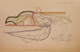
Show 974 MRS. O. A. MERRITT HAWKES OX THE [Dec. 11, that canal, then this ramus in Chlamydoselachus is equivalent to the true ramus oticus plus a few more nerve-fibres. The r. oticus does not supply all the sense-organs which occur in the region of the canal anterior to the commissural canal, but apparently two of these are supplied by a dorsal branch of the Glosso-pharyngeus and the remainder by a, dorsal branch of the Yagus. The branches of the ramus buccalis are arranged approximately in two groups : one group being associated with the maxillary branches of the mandibularis Y.,and the other with the branches of the maxillaris Y. The branches not only supply the diffuse group of ampullae which occurs above the mouth, but also a, group which lies between the descending portion of the infra-orbital canal and the spiracle. This group is not usually represented in Elasmobranclis. It is noteworthy that each of the branches supplies both ampullae of Lorenzini and neuromasts. This fact, and the diffuse condition of the buccalis branches seem to indicate that the nerve under discussion is in a primitive condition. The anastomoses, which are so conspicuous in dissecting the branches of the buccalis, indicate the first step from a primal to the secondary condition, such as is found in most Elasmobranclis, where the single buccalis of this species is represented b}~ two or three main branches. There are similar anastomoses in that portion of the external mandibularis YII. where it is at some distance from the neuromasts supplied. The anastomoses in the case both of the buccalis and external mandibularis YII. indicate a secondary simplification of the nerve-supplv. (c) The truncus hyomandibular is of the Facialis originates from the medulla by two roots at the same level as the roots of Y. and YII. It passes at first into the large foramen common to the whole trigemino-facial complex, then bends backward and passes outward through its own foramen. The distal end of the cephalad root, which is probably the external mandibular lateral line root, is connected with the Gasserian ganglion by several small strands- the rami communicantes (Plate LXYIII. fig. 2, R.C.): posteriorly, the complex hyomandibular ganglion is connected with the auditory nerve by the pars intermedia (fig. 2, P.I.). General cutaneous fibres enter the hyomandibular ganglion from the Gasserian ganglion by the rami communicantes. About 5 mm. from the brain, just as they emerge from the chondrocranium, the hyomandibular roots unite and swell into a large ganglion, bilobed at its distal extremity. The cephalad portion-the geniculate ganglion-gives rise to the pre- and post-trematic YII. rami (fig. 1, Pr.F. & Pt.F.). The caudad portion, which is the smaller, is the external mandibular ganglion-that is, the most caudad lateralis ganglion of the Facialis. This ganglion gives rise to the truncus hyomandibularis, which contains the lateralis components which separate out as the rami externi mandibulares, as well as some components of a brancliiomeric nerve. There may be, in a form as low as Chlamydoselachus, general cutaneous fibres belonging properly to the Facialis root, as well as |