OCR Text |
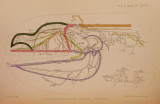
Show 1906.] OF SOUTHERN INDIA AND CEYLON. 665 an additional reason for pulling tlie alimentary tube back to give more room. A further examination by sections of Doridopsis reticulata (Cockerell & Eliot, J. of Mai. xii. 1905, p. 41) indicates that this species also is a Doriopsilla. The real buccal ganglia are not, as stated, 4 mm. behind the central nervous system, but close to it and united by short connectives to the part that seems to correspond to the pleural ganglia. The alimentary tube differs somewhat from that of D. reticulata. The part in front of the nerve-collar is broad and pouch-like : just under the collar (as preserved) is a valvular apparatus separating this anterior dilated portion from the long narrow posterior portion. Before entering the liver the alimentary tube dilates again into a pouch divided by a constriction. Under the buccal parts is a large bilobed gland, probably, but not demonstrably, connected with the proboscis. D o r io p s il l a m in ia t a (A. & H.). (Plate XLVII. figs. 4 & 6.) (A. & H. I.e. p. 130.) Four specimens are preserved, hardened and in bad condition, but quite recognisable. The following notes are the result of examining recent specimens from Karachi, ~ which I have no hesitation in identifying with this form. The animal was described by me in the ‘ Journal of Conchology,' vol. ii. no. 8, Oct. 1905, as Doridopsis miniata, and stated to have long buccal connectives. Subsequent examination by sections has shown that this is incorrect. The portion of the alimentary canal lying between the central nervous system and the liver is supported by several strong filaments, and some of these were mistaken for connectives running between the buccal ganglia and the central nervous system. The sections show clearly that the ganglia are arranged in the manner characteristic of Doriopsilla. The main mass (a) is of somewhat irregular shape and surrounds the oesophagus. Immediately behind it and touching it, but below the oesophagus, are the two buccal ganglia close to one another. Not far from the buccal ganglia a short connective runs to the right and terminates in a small ganglion near the penis, which apparently innervates the genitalia. The anterior portion of the alimentary canal is a thin tube, which preserves a uniform diameter until it enters the liver. On issuing from the proboscis it describes a Z-shaped figure. The lower curve of this figure is free and lies below the nerve-collar, which surrounds the upper loop. On reaching the liver the tube runs for some distance just below the surface before descending into the interior. In spite of a careful examination, the presence of ptyaline or salivary glands could not be demonstrated. It seemed certain that the wall of the proboscis does not contain a long duct coming from the ptyaline gland as in Doridopsis nigra. |