| OCR Text |
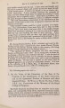
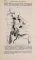
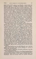
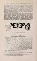
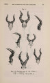
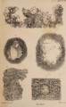
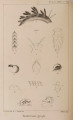
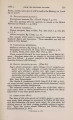
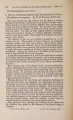
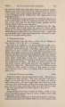
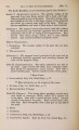
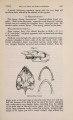
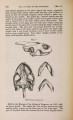
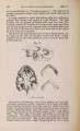
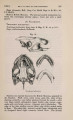
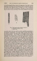
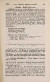
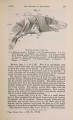
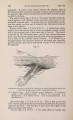
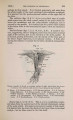
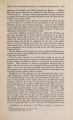
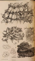
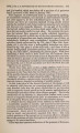
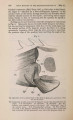
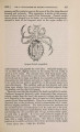
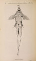
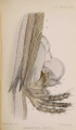
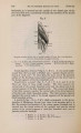
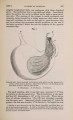
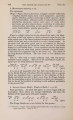
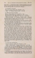
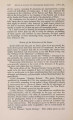
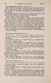
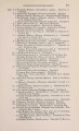
Show 462 MR. ST. GEORGE MIVART O N T H E [June 24, also goes to the digits, but which takes origin from the distal half of the ulna. Pelvic Limb. Semimembranosus (figs. 8, 9, 10, & 11, S.M). This slender muscle springs from the hypapophyses of the caudal vertebrae (the first two that are elongated), and passing forwards, enclosed in a sheath consisting of the subcaudal muscular mass, joins the posterior margin of the gracilis and origin of the semitendinosus. It passes along external to the large precloacal glandular mass, and side by side with similarly directed and similar-sized femoro-caudal and ischio-caudal. Fig. 8. Superficial muscles of ventral surface of right side. A. Adductor. Ex. O. External oblique. F. C. Femoro-caudal. E. B. Flexor digitorum. F. H. Flexor hallucis. G. Gracilis. I. Iliacus. /. C. Ischio-caudal. S. Sartorius. S. M. Semimembranosus. S. T. Semitendinosus. The ischio-caudal (figs. 8 & 11, I.C) is the most internal of the caudal appendicular muscles. It arises from the subcaudal muscular mass at the same distance backwards as does the semimembranosus ; passing forwards, it is inserted into the posteroexternal angle of the ischium. Femoro-caudal (figs. 8, 9, 10, & 11, F. C). This muscle is about the same size as that last described, and also springs from the anterior caudal hypapophyses. It comes out of the sheath formed for it and the muscles running parallel with it by the subcaudal |