| OCR Text |
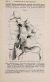
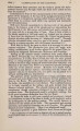
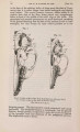
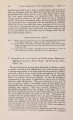
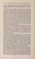
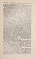
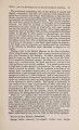
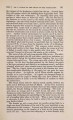
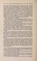
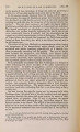
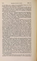
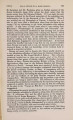
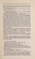
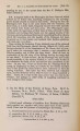
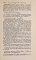
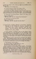
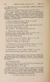
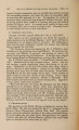
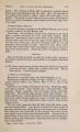
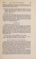
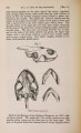
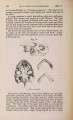
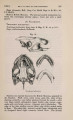
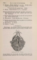
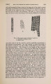
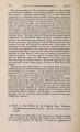
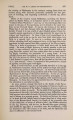
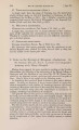
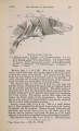
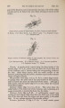
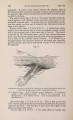
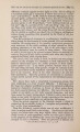
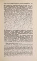
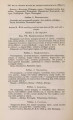
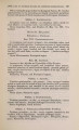
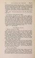
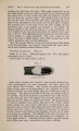
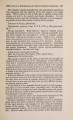
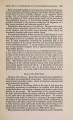
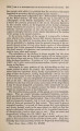
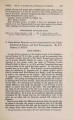
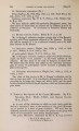
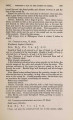
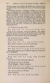
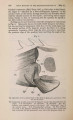
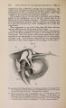
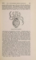
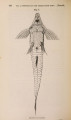
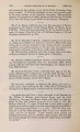
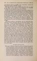
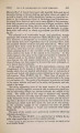
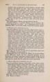
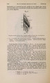
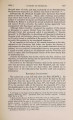
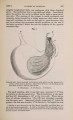
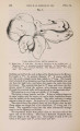
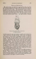
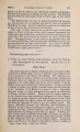
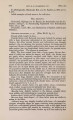
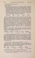
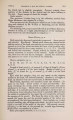
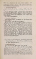
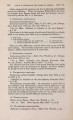
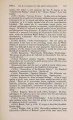
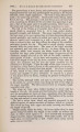
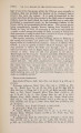
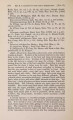
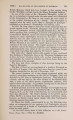
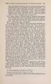
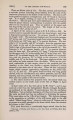
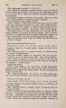
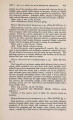
Show 1869.] MYOLOGY OF MENOBRANCHUS LATERALIS. 465 and size to the gluteus maximus. Arising from the outer side of the ilium, it passes down beside the last-named muscle, and is inserted by aponeurosis into the inner side of the upper part of the tibia. Gluteus medius (?) (fig. 10, G.Md). Arising from the front of the ilium, this small muscle passes down beside the iliacus, and is inserted into the upper part of the outer side of the femur. Gluteus minimus (?) (fig. 10, G. Mi). This little muscular bundle passes from the posterior side of the ilium to the upper half of the posterior side of the shaft of the femur. Fig. 11. --7J Deeper muscles of ventral or flexor surface of pelvic limbs, the gracilis, semitendinosus, and semimembranosus being cut and reflected on the right side, and the adductor also on the left side. A. Adductor. B. Biceps. Ex. 0. External oblique. F. C. Femoro-caudal. F. B. Flexor digitorum. F. H. Flexor hallucis. G. Gracilis. I. C. Ischio-caudal. I. F. Ischio-femoral. /. P. Ilio-peroneal. S. Sartorius. S. M. Semimembranosus. S. T Semitendinosus. Ischio-femoral (fig. 11,1. F). A very small muscle (which m ay perhaps answer to the quadratus femoris of higher animals) passes from the postero-external angle of the ischium to the head of the femur very near the acetabulum. Ilio-peroneal (figs. 10 & 11, I. P). A very long and slender muscle arises from the ilium, immediately beneath and closely connected with the gluteus maximus. It is inserted into the peroneal side of the fibula above its middle. The biceps (?) (fig. 11, B) is also a very slender muscle. It extends from the shaft of the femur, just below the insertion of the femoro-caudal, downwards to the lower part of the fibula. Tibialis anticus (figs. 9 & 10', T. A and T. Al). This muscle seems to be double in Menobranchus. It arises from the front of the distal end of the femur, and from the proximal parts of the tibia |