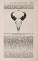
| OCR Text |
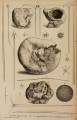
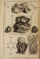
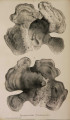
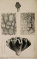
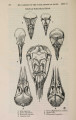
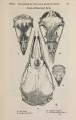
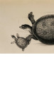
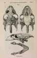
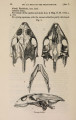
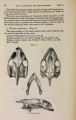
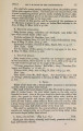
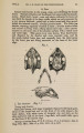
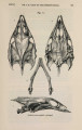
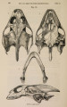
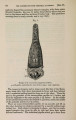
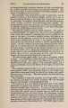
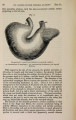
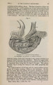
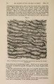
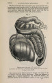
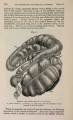
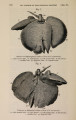
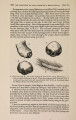
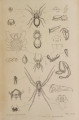
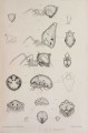
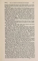
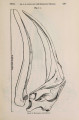
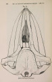
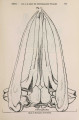
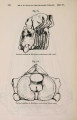
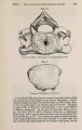
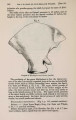
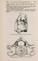
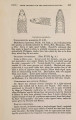
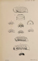
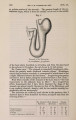
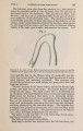
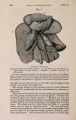
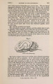
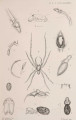
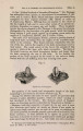
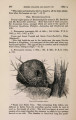
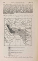
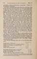
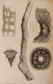
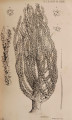
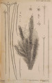
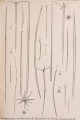
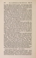
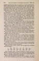
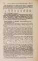
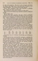
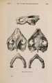
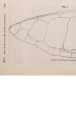
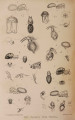
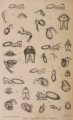
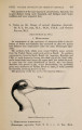
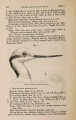
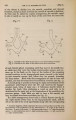
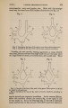
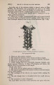
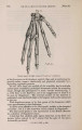
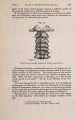
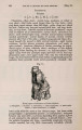
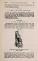
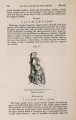
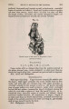
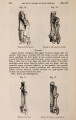
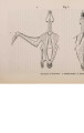
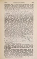
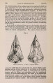
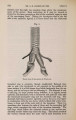
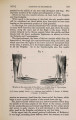
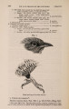
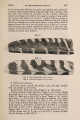
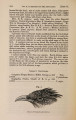
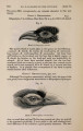
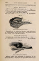
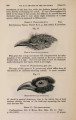
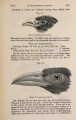
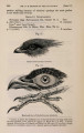
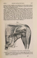
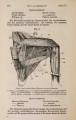
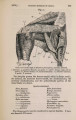
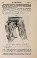
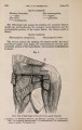
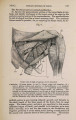
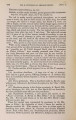
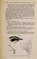
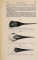
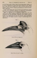
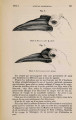
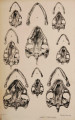
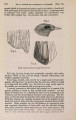
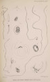
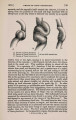
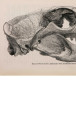
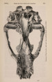
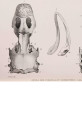
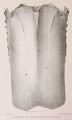
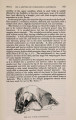
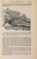
Show 1873.] DR. E. L. MOSS ON A VIRGULARIAN ACTINOZOON. 731 The specimen from which the following notes are made has indeed already suffered considerable post-mortem disintegration; its more delicate portions do not admit of the handling necessary for dissection under the microscope. Sufficient, however, remains to enable m e to speak definitely as to its position in the animal kingdom, though the absence of means of reference in this colony makes complete accuracy unattainable. The creature belongs to the Virgularian section of the family Pennatulidae, but differs from those of its genus that I know of in that the lateral ridge-like processes bearing the polypes exist only on one side of the central axis ; in short, to borrow a term from its fossil relatives the Graptolites, it is " monoprionian." In this example the pale rosy chocolate tint, so common in its class, is still preserved; the entire actinosoma (Plate L X I . fig. 1) measures 8 feet 6 inches in length. The lower or proximal foot-length (A) is the thickest part; its diameter is a little over 1 inch; it is cylindrical; its end tapers rapidly to a soft cone perforated by a pore (fig. 2) ; and it is grooved externally by twenty-four longitudinal wrinkles. This lower foot-length consists of two cylinders of cartilaginous consistence, the inner, more transparent and firmer, closely enclosing the central hard rod or coral, the outer investing the former and attached to it only by four thin longitudinal septa (fig. 3), passing through the entire thickness of the inner cylinder, but very slightly attached to the central rod. The space included between the two concentric cylinders is thus divided into four equal chambers, ending below at the apex, and above gradually narrowed by the widening of the partitions till they finally disappear about 18 inches from the proximal extremity. A section of this part presents the wheel-like appearance shown in fig. 3. Externally a line of minute pores marks the position of each septum ; but I could not trace any connexion between them and the quadrate chambers. The second foot-length of the actinosoma is m u c h more slender (fig. 1, B ) ; the sarcode closely invests the central axis and presents a twisted appearance. The four lines of pores are still visible externally ; but the space between the layers of soft tissue is greatly reduced, and at the distal end of this length terminates altogether. At this part the two lines of pores on one half of the organism change their character; the openings, instead of being very minute and isolated, become larger, and a little further on begin to occur in rows of twos and threes. The fleshy parts here project more from the central axis on this side than on the other. At the end of another foot-length the pores have changed into little rows of pits on either side of a ridge of soft tissue, each pit filled with its polype ; and from this part upwards the polype-bearing part of the ccenosarc (fig. 1, C ) extends to a gracefully tapering extremity, but presents an unchanged plan of structure throughout. Taking a central part for example (fig. 4), w e find the hard round axis of very much the same diameter as below, though above it tapers with the rest of the tissues till it ends in a fine point. It is very thinly covered by the two fleshy layers for about two thirds of its circumference (section, fig. 5 ) |