| OCR Text |
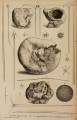
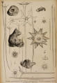
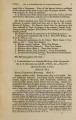
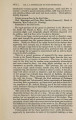
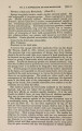
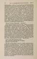
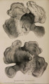
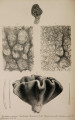
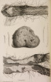
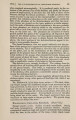
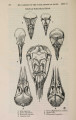
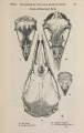
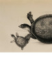
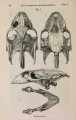
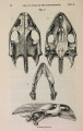
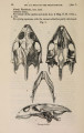
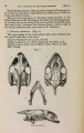
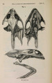
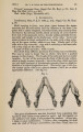
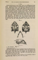
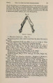
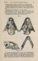
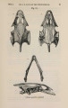
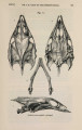
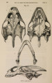
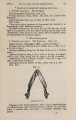
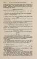
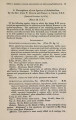
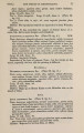
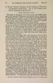
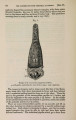
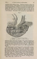
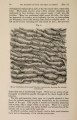
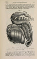
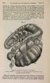
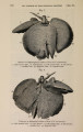
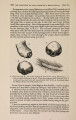
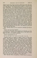
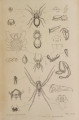
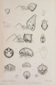
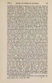
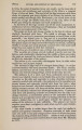
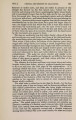
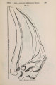
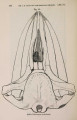
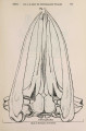
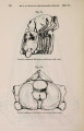
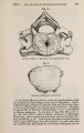
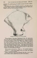
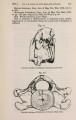
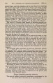
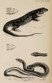
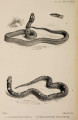
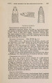
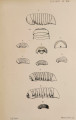
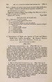
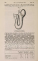
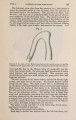
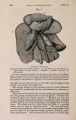
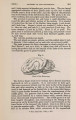
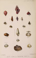
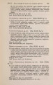
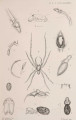
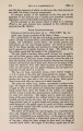
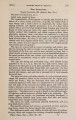
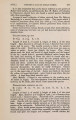
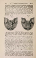
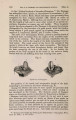
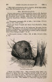
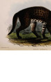
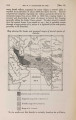
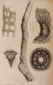
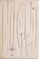
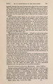
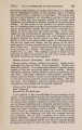
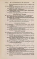
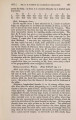
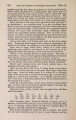
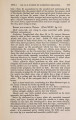
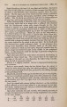
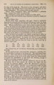
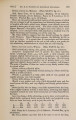
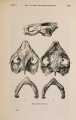
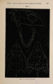
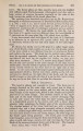
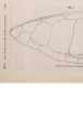
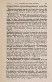
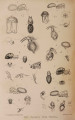
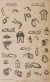
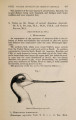
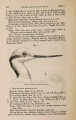
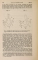
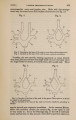
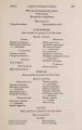
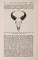
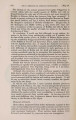
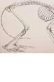
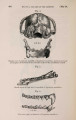
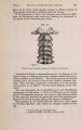
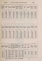
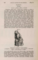
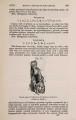
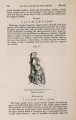
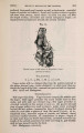
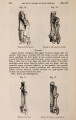
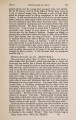
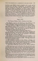
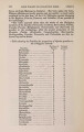
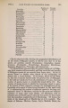
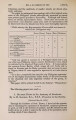
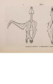
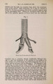
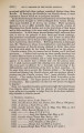
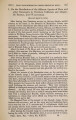
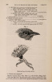
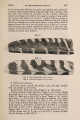
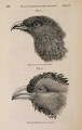
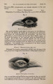
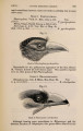
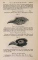
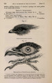
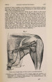
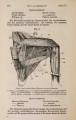
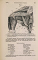
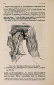
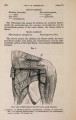
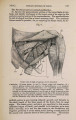
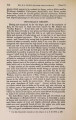
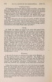
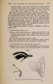
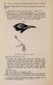
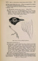
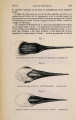
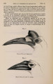
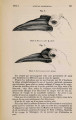
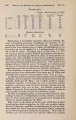
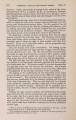
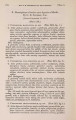
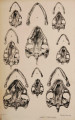
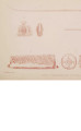
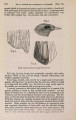
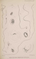
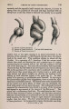
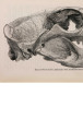
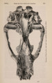
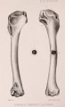
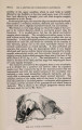
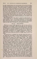
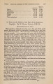
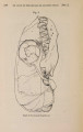
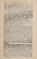
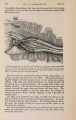
Show 740 DR. T. s. COBBOLD ON N E W OR RARE ENTOZOA. [NOV. 18, me that Dr. Little, of Shanghai, is diligently engaged in working out the structure and development of the parasite. Dr. Krabbe, of Copenhagen, has likewise supplied some interesting particulars; but, in the absence of any references, I a m led to conclude that the Danish author's observations are based on the previously published statements of Prof. Leidy (" Husdyrenes Indvoldsorme," Tidsskrift for Vet. 2den Rsekke, ii. 1872). O n the the 15th of April last I examined a number of these worms, two of which, male and female, are represented in the accompanying Plate. To the naked eye the sexual differences are readily discernible. The female (fig. 5) maintains almost throughout a uniform calibre of about ^ inch ; but at the head it diminishes to -M. inch, and at the tail to about T±-$ inch, the caudal point being bluntly convex. The oviducts of all the females examined were crowded with eggs, and in certain situations the eggs were collected together in the form of large ovoid masses. The largest eggs had a long diameter o f - ^ inch, with an average breadth of -4Vo incn. These contained coiled embryos; the diameter of their bodies varying from 10-\nr inch to ygta inch. _ The male parasite is readily recognized by its comparatively slender body, having a diameter of about £0- inch, and also by its elegant spirally curved tail, which is three or four times twisted upon itself with the regularity of a corkscrew (fig. 6). The coiled portion is much narrower than the body of the worm, and it finally dwindles down to a breadth of -jJ^ inch, its extreme point being blunt, as in the female. Within a short distance of the extremity the two spicules, of unequal length, m a y often be seen projecting from the cloacal outlet, this part of the worm being also furnished with a well-marked horseshoe-shaped bursa (fig. 7). This organ may be described as consisting of two transparent folds or extensions of the cuticle, each lateral division of the hood being supported by four oval glandular rays. The rays are apparently eight in number, and arranged in pairs. The uppermost pair is the largest, the other pairs gradually decreasing in size from above downwards. When viewed laterally, these oval rays present a beaded appearance, collectively forming a rather striking microscopic object. The diameter of the largest ray is only about the y ^ - inch, whilst that of the smallest is not more than y-g-W in c n fr°m side to side*. As seen in the drawing, the epidermal layer of the skin was generally found projecting more or less beyond the limit of the dermis. I regarded this as a post mortem production. In one case * Since this paper was read to the Society m y attention has been called to a much more detailed description of the worm by Mr. Welch, of Netley, communicated to the ' Monthly Microscopical Journal' for October 1873, p. 157. The author gives many particulars that have escaped m y notice, and he also interprets some of the facts observed by us both in a very different sense from that I have adopted. He recognizes but one spiculum, and regards the oval rays as generative appendages, of the nature of vesiculee seminales, communicating with the seminal duct. He observed twelve of these oval appendages, and, amongst other things, describes the intestinal tube as terminating in a blind caecal extremity.- T. S. C. |