| OCR Text |
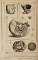
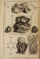
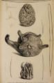
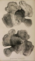
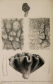
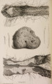
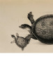
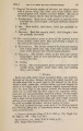
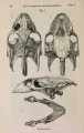
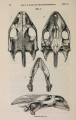
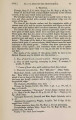
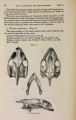
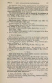
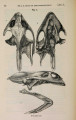
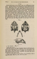
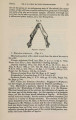
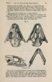
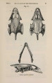
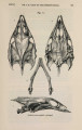
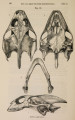
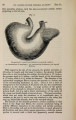
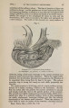
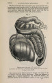
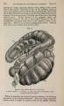
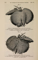
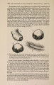
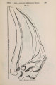
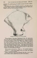
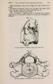
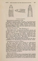
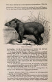
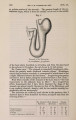
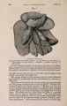
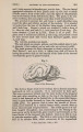
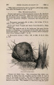
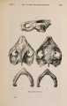
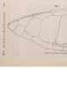
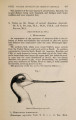
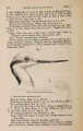
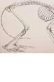
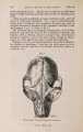
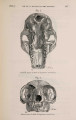
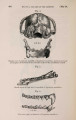
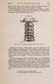
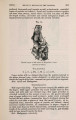
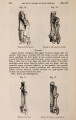
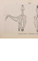
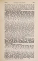
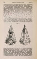
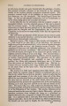
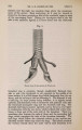
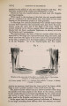
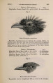
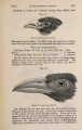
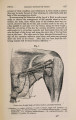
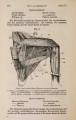
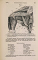
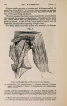
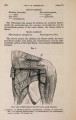
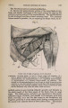
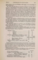
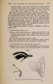
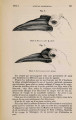
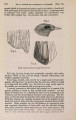
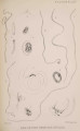
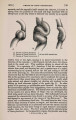
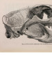
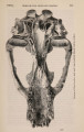
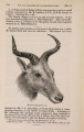
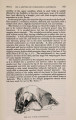
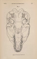
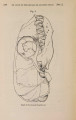
Show 202 MR. GARROD ON THE DEATH OF A KANGAROO, [Feb. 18, fissure; it embraces the middle gyrus, and does not cease opposite its posterior superior angle, but descends about halfway down its posterior limb to end by a point. The whole brain narrows in front; and the crucial sulcus is not at all strongly marked. The corpora albicantia are separated behind ; and the optic nerves in front of the chiasma run forwards close together. The pituitary body is of fair size. 2. On the Cause of Death of a Black-faced Kangaroo (Macropus melanops). By A. H . G A R R O D , B.A., F.Z.S., Prosector to the Society. tk [Received February 18, 1873.] The cold weather of the first week of this month coming on rather suddenly, seems to have been the cause of the death of three animals in the Gardens, in all of which, on post mortem examination, it was found that the lesion was the result of excessive and abnormal movement in the abdominal viscera. A Paradoxure died from intussusception of the small intestine, part going through the ilio-caecal valve into the colon; an E m u from prolapse of a considerable length of the alimentary canal; and the above-named Kangaroo from strangulation of a loop of small intestine by the tight twisting round it of the caecum-a most uncommon lesion, which proves that the possession of that appendage has its disadvantages as far as the individual is concerned-just as in several human subjects death has been proved to have occurred from impaction of small bodies, like cherry-stones, in the appendix vermiformis. In the Kangaroo under consideration, on opening the abdomen the attention was immediately drawn to a large loop of strangulated small intestine, quite black from congestion, and partly covered with flakes of recent lymph, the result of the induced peritonitis, which was inconsiderable. The length of gut involved was nearly two yards after it had been detached from the mesentery; but in the body of the animal it appeared considerably shorter, from being convoluted in the ordinary manner. The last foot or so of the small intestine was not included in the diseased loop, which consisted of the portion immediately preceding it. The caecum was about a foot and a half long, and was situate in the right iliac region, from which it extended to the left superficially, and then again to the right behind the loop of intestine which it encircled, so that the caput caeci could be seen, distended with grumous matter (as was the strangulated portion), to the right. With care, while the viscera were in situ, the little finger could be introduced into the ring thus artificially formed ; and it was evident that the constriction was mostly produced by the mesenteric band which attaches the proximal portion of the caecum to the small intestine. There were no adhesions of importance. The viscera were removed en masse; and afterwards, without the least difficulty, the caecum was uncoiled, and the intestine was then left quite per- |