| OCR Text |
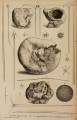
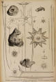
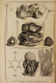
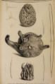
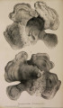
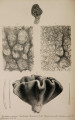
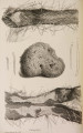
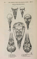
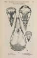
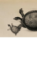
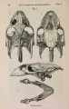
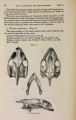
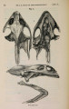
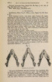
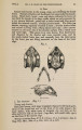
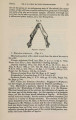
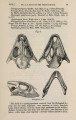
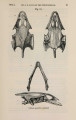
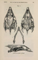
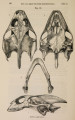
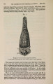
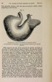
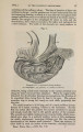
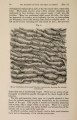
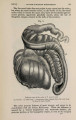
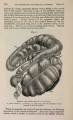
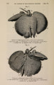
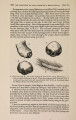
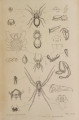
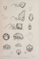
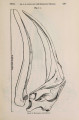
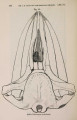
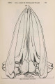
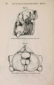
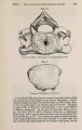
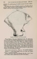
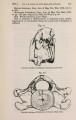
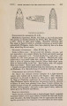
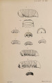
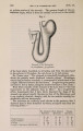
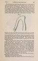
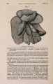
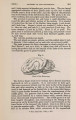
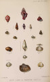
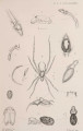
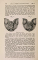
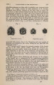
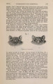
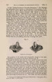
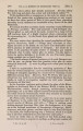
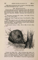
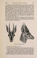
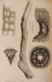
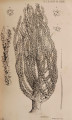
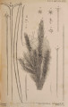
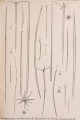
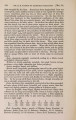
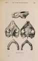
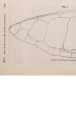
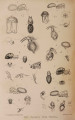
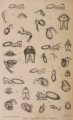
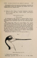
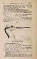
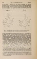
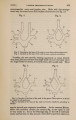
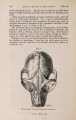
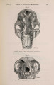
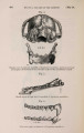
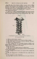
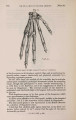
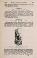
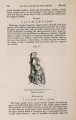
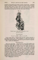
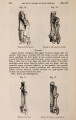
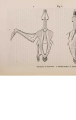
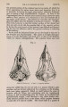
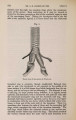
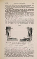
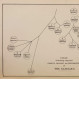
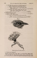
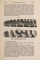
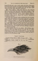
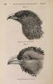
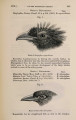
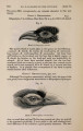
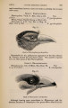
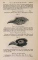
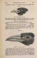
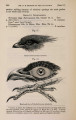
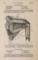
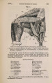
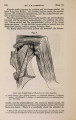
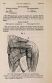
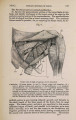
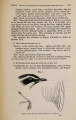
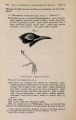
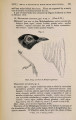
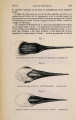
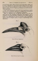
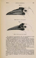
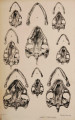
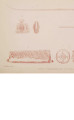
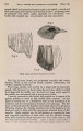
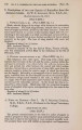
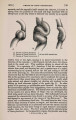
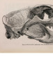
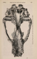
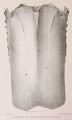
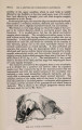
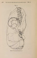
Show 24 DR. J. S. BOWERBANK ON THE SPONGIADcE. [Jan. 7, Fig. 3. An attenuato-patenti-ternate connecting spiculum of about the average size, magnified 80 linear. Fig. 4. The head of one of the largest and most fully developed connecting spicula, magnified 80 linear. Fig. 5. One of the attenuato-stellate retentive spicula from the interstitial membranes, magnified 530 linear. Tethea simillima, Bowerbank. Fig. 6 represents the type specimen in spirit in the Museum of the Koyal College of Surgeons, London: natural size. Fig. 7. The small specimen in the dried state, exhibiting a view of the external surface, natural size. Fig. 8. A sectional view of the same specimen that is represented by figure 7, showing the central nucleus and the mode of disposition of the skeleton-fasciculi : natural size. Fig. 9. T w o thirds of one of the large fusiformi-acerate skeleton-spicula, magnified 80 linear. This figure also represents the same form of spiculum as an external defensive one. Fig. 10. One of the fusiformi-porrecto-ternate external defensive spicula, magnified 80 linear. Fig. 11. A n attenuato-recurvo-ternate defensive spiculum, with long and very slender shaft, magnified 80 linear. Fig. 12. One of the stout fusiformi-acerate spicula that surround the defensive fasciculi of the external surface, magnified 80 linear. Fig. 13. A small portion of the skeleton of one of the gemmules of the sponge, extending from its centre to its external surface, shewing its unihamate and porrecto-ternate spicula in situ, from a specimen mounted in Canada balsam : magnified 80 linear. Tethea Cliftoni, Bowerbank. Fig. 14. The type specimen, showing the remarkable mode of its location under difficulties: natural size. Fig. 15. One of the fusiformi-acuate skeleton-spicula, magnified 150 linear. Fig. 16. One of the large sphero-stellate spicula of the dermal rind of the sponge, magnified 530 linear. Fig. 17. A minute subsphero-stellate retentive spiculum with clavated cylindrical radii, magnified 530 linear. Fig. 18. A smaller specimen of the same description as that represented by fig. 17, magnified 530 linear. PLATE IV. Leuconia glomcrosa, Bowerbank. Fig. 1. The type specimen, natural size. Fig. 2. One of the equiangular triradiate spicula of the dermal membrane, magnified 80 linear. Fig. 3. One of the largest-sized fusiformi-acerate dermal spicula, which bas been fractured near its middle and cemented together again: magnified 80 linear. Fig. 4. A small-sized fusiformi-acerate dermal spiculum, magnified 80 linear. Figs. 5 & 6. T w o of the triradiate spicula of the interstitial skeleton, magnified 80 linear. Desmacidon fistulosa, Bowerbank. Fig. 7 represents the type specimen, natural size. l^g. 8. One of the skeleton-spicula, magnified 150 linear. Ciocalypta Tyleri, Bowerbank. Fig. 9. The type specimen, natural size. |