| OCR Text |
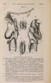
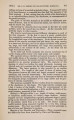
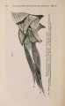
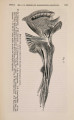
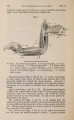
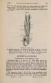
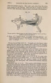
Show 1870.] MYOLOGY OF PLATYDACTYLUS JAPONICUS. 421 pubis, and in front to the sides of the rectus abdominis; its fibres are directed from behind forward and towards the dorsal surface. Intercostals are the layer of muscles next internal to the last; they are united behind with the internal oblique and transversalis to form an arch over the external and back part of the thigh ; they occupy the spaces between all the ribs from the sides of the rectus and sternum to the vertebrae, anteriorly they are continuous with the scalenus ; their fibres are directed forward and slightly towards the dorsal surface. Internal oblique and transversalis are situated between the ribs and the viscera. Posteriorly they are united together and to the intercostals as above mentioned; dorsad they are attached to the angles of the ribs internally, at the place where the external oblique is attached externally; ventrad they are attached to the side of the rectus abdominis, and, in front of that, to the end of the fourth sternal rib, to the xiphisternum, and to the postero-lateral edge of the sternum. Anteriorly they extend as far as the fourth cervical rib. The fibres of the internal oblique are directed obliquely forwards and towards the ventral surface ; those of the transversalis go transversely across the body. Quadratus lumborum arises from the anterior edge of the sacrum. It is attached to the sides of six lumbar and the last dorsal vertebrae, and encloses all the lumbar ribs in a thick muscular mass, which terminates anteriorly at the last dorsal rib. Retrahentes costarum line the whole dorsal surface of the abdominal cavity in front of last muscle. They consist of muscular slips separable by dissection, each arising from a centrum of a vertebra, and passing ventrad of the rib of its own vertebra ; and that of the next one in front is inserted into the next but one, interdigitating with the transversalis and internal oblique; the most anterior one is attached to the centrum of the first dorsal vertebra and to the fourth cervical rib, and the last to the twelfth dorsal. Sartorius occupies the greater part of the ventral aspect of the thigh. It arises from the hook-like process of the pubis in front of the acetabulum, also from the whole length of the ilio-pubic ligament ; it has a broad muscular insertion into the lower or inner side of the tibia, occupying one quarter of its proximal end. Gracilis arises from the side of the ischial symphysis and from the ventral part of a tendinous intersection which connects the posterior point of that bone with the posterior end of the ilium, and which seems to represent the tuber ischii; it is inserted partly tendinous and partly fleshy into the tibia, beneath the last muscle. Transversus perinei is a small muscle in front of the cloaca, arising from the proximal side of the above-mentioned tendinous intersection, and is inserted into a cartilaginous rod attached to the posterior end of the ischium, and into the skin anterior to the cloaca. Semimembranosus arises from the tendinous intersection dorsad of the gracilis, and is inserted into the tibia on the inner side of the internal lateral ligament, and nearer the head of the bone than the gracilis. There are two separate muscles which, perhaps, represent |