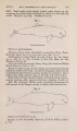
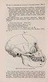
| OCR Text |
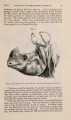
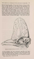
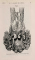
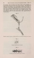
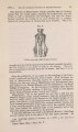
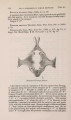
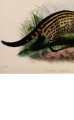
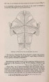
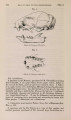
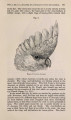
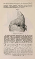
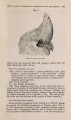
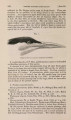
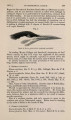
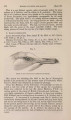
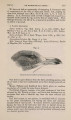
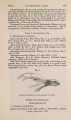
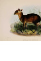
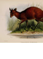
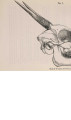
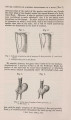
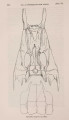
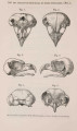
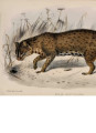
Show 1871.J MYOLOGY OF THE KINKAJOU. 557 ratores externus and internus, and gemelli. They present the same attachments and disposition as the corresponding muscles in man. The gluteus maximus is a large, broad, and somewhat extensive muscle. It arises from the upper and posterior aspect of the iliac crest, from the sacral aponeurosis which covers the sacro-caudal muscles, and from the transverse processes of the third and fourth caudal vertebrae. It is inserted into the posterior aspect of the shaft of the femur, occupying fully its middle three-eighths. Its upper fibres only are tendinous at their insertion. The gluteus medius arises from the upper two-thirds of the dorsal surface of the ilium, from the aponeurosis of the gluteus maximus, and also that separating it from the sacro-caudal muscles; it is more or less continuous with the pyriformis muscle, and inserted along with it into the great trochanter of the femur. The gluteus minimus preserves its usual arrangement; the gemelli and obturator internus muscles seem to be differentiations from it. The tensor fascice femoris is a fusiform muscle. It arises from the ilium (below the sartorius) by a pointed tendon; the muscle is about an inch and a quarter long, and terminates in the fascia of the thigh. The capsular ligament of the hip-joint is very strong; the ligamentum teres only moderately so. The gastrocnemius has a sesamoid bone developed on its outer head ; it presents no decidedly interesting peculiarities. The soleus is a single-headed muscle. It arises from the head of the fibula, and from the peroneal intermuscular septum. It is inserted into the os calcis along with the preceding. The plantaris is a very large muscle. It arises from the outer femoral condyle, and is also attached to the sesamoid bone belonging to the outer head of the gastrocnemius. It terminates in a strong tendon which traverses the inner aspect of the os calcis, and, becoming expanded in the sole of the foot, forms the plantar fascia. It is closely associated in the sole of the foot with the flexor brevis digitorum. In the Caracal, Dog, and Paradoxurus this muscle is not so large. In other respects it does not materially differ from that of the Kinkajou. The popliteus muscle is very large and fleshy. The anterior tibial artery passes above its upper instead of below its lower border as in the human subject. Occasionally, however, this peculiar mode of distribution of the artery is met with in man; I met with one instance of it during the last winter session. The abductor minimi digiti is aborted at the sixth metatarsal base, constituting Wood's abductor ossis metatarsi quinti. The flexor brevis digitorum pedis is distributed only to the second, third, and fourth digits; each tendon, prior to its splitting for the passage of the perforans, is joined by a fleshy slip from the accessorius, given off from the latter opposite the point of junction of the long flexors with the accessorius. The perforatus tendon of the fifth digit is derived from a distinct wedge-shaped muscle, which springs |