| OCR Text |
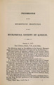
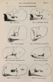
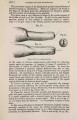
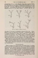
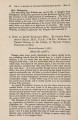
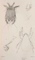
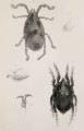
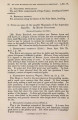
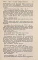
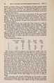
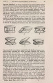
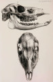
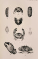
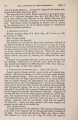
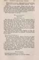
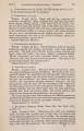
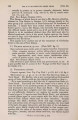
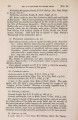
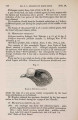
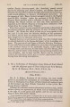
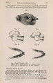
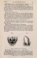
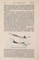
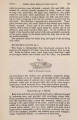
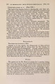
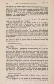
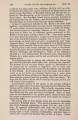
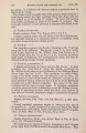
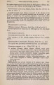
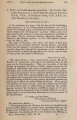
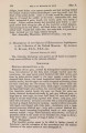
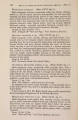
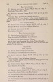
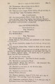
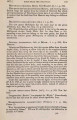
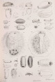
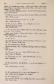
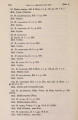
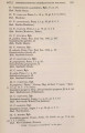
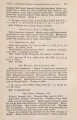
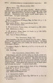
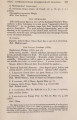
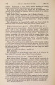
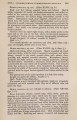
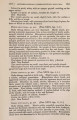
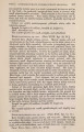
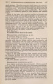
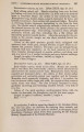
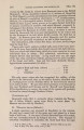
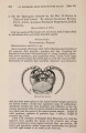
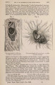
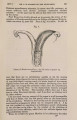
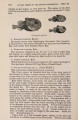
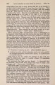
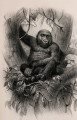
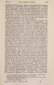
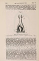
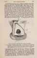
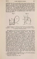
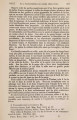
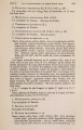
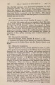
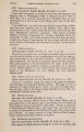
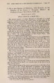
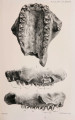
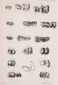
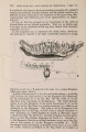
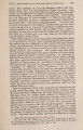
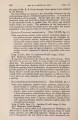
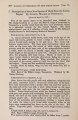
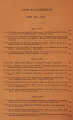
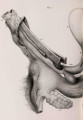
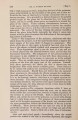
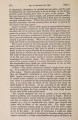
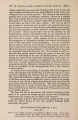
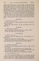
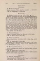
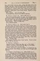
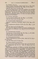
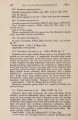
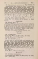
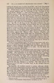
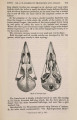
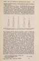
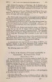
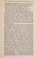
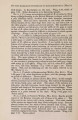
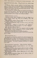
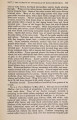
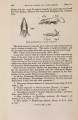
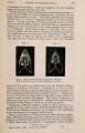
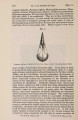
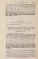
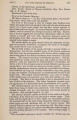
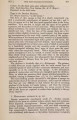
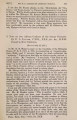
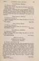
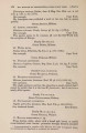
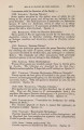
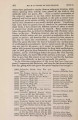
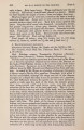
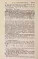
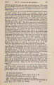
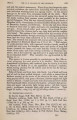
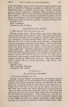
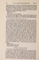
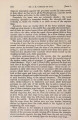
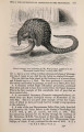
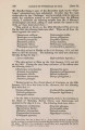
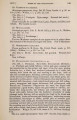
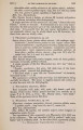
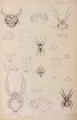
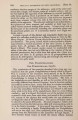
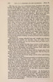
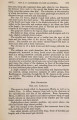
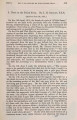
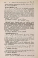
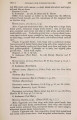
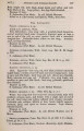
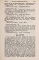
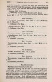
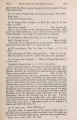
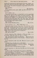
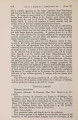
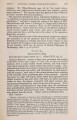
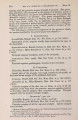
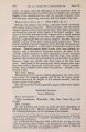
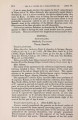
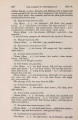
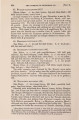
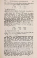
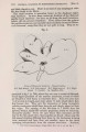
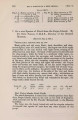
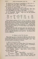
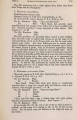
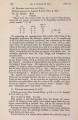
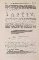
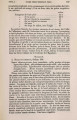
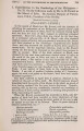
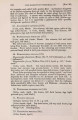
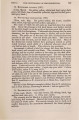
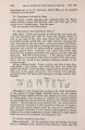
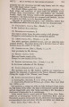
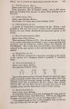
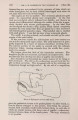
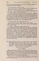
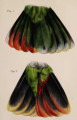
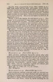
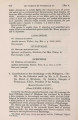
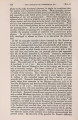
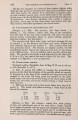
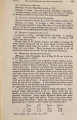
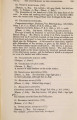
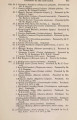
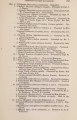
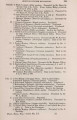
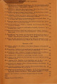
Show 710 VISCERAL ANATOMY OF RHINOCEROS SONDAICUS. [Nov. 6, and thick digestive coat. There is no trace of any oesophageal valve like that found in the Horse. The small intestine is somewhat larger in the duodenal region than elsewhere. Its fir3t three inches are destitute of the flattened papillae found elsewhere; but here, as all along the small intestines, minute villi are present everywhere. Three inches from the pylorus the papillee commence, and resemble those similarly situated in Rhinoceros unicornis1, except that they are not quite so long. They are re- Fig. 3. Liver of Rhinoceros sondaicus. Visceral surface. L.L. Left lateral. L.C. Left central. B.C. Eight central. B.L. Right lateral. C. Caudate. Sp. Spigelian lobe. presented in fig. 1 (p. 708), where they are seen to consist of flattened, round-tipped processes of the mucous membrane, several of which are blended at their bases, in transverse lines. None are more than *3 of an inch in length, and most about *6 inch broad where they first become free. They give the impression of being incomplete valvulae conniventes which have been cut and deeply jagged at their free edges. The opening of the bile-duct is 7 inches from the pylorus, being a nipple-like tubular projection, nearly an inch long, among the papillae. From the spot where they commence, all the way to the ileo-caecal valve, these papillae are found-those near the last-named situation differing from those in the duodenum in being more scattered and freer from one another, many in the ileum springing independently from the mucous membrane. Nowhere, 1 Vide Prof. Owen's figure, Trans. Zool. Soc. vol. iv. pl. xii. fig. 1. |