| OCR Text |
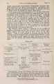
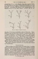
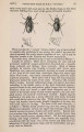
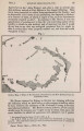
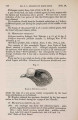
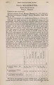
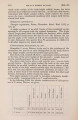
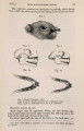
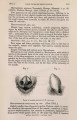
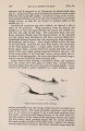
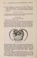
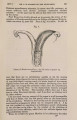
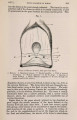
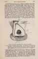
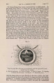
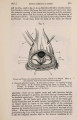
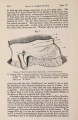
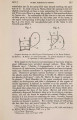
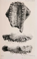
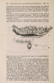
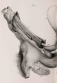
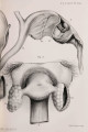
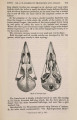
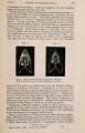
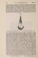
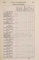
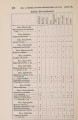
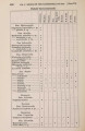
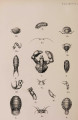
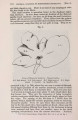
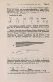
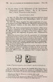
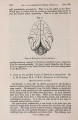
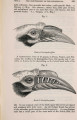
Show 1877.] ANATOMY OF HYJENA CROCUTA. 373 the peritoneal ovarian pouch, and is provided with numerous elongated fimbriae. From this extremity the tube passes downwards lying in the anterior wall of the sac as far as its floor, where it turns upward and then comes to lie in the posterior wall of the pouch, terminating by becoming continuous with the corresponding uterine horn at the point of attachment to the latter of the ovarian ligament. The tube thus accommodates within the concavity of its flexure the fundus of the ovarian pouch. Uterus.-The uterus consists of a central body, and two horns. Each horn measures 3| inches in length, and extends from the uterine ligament of the ovary as far as the middle line, where it meets with its fellow to form the corpus uteri. In this course it is contained between the folds of the broad ligament, and increases gradually in calibre from without inwards. Midway between its commencement and termination there is attached to the horn a stout fibrous cord, which evidently represents the round ligament of the uterus. When traced backwards it is seen to enter the inguinal region; but its exact attachment in this locality I could not determine, in consequence of the organs having been removed from the body before I was aware that the generative organs of this Hyaena presented any unusual arrangement. At the point of attachment of the round ligament to the left uterine horn there was a small pedunculated hydatid. The body of the uterus is formed by the junction of the right and left horns, and lies between the bladder and the rectum. It measures 5 inches in length from the junction of the horns to the opening of the os uteri into the urino-genital canal. That the whole of this is to be regarded as corpus uteri, and not as constituting any part of the vagina, is proved by the absence of any constriction in its interior which might correspond to an os uteri, the tubular body of the uterus remaining of the same calibre, and having the walls of uniform thickness down to its opening into the urino-genital canal. What must, therefore, be regarded as the os uteri is the constricted opening by means of which the uterus communicates with the canal common to both urinary and generative organs. At this point there is a thick semilunar fold of mucous membrane, the concavity of which is directed backwards, and its margins attached to the walls of the urinogenital canal. It is placed horizontally, having its surfaces directed upward and downward, and intervenes between the opening of the urethra above and that of the os uteri below it into the common canal. Internal to this fold is the uterus ; external to it is the urino-genital canal • for, as we shall see presently, there is in this animal a complete absence of any differentiation of vagina from urethra. The mucous lining of the uterus is thrown into weil-marked longitudinal rugse. Urino-genital Canal.-As already stated, the uterus and urethra open into a common canal. This, the urino-genital canal, extends from the junction of the two tubes to the extremity of the clitoris, and measures rather more than 8 inches. In this course it describes a wide curve with the convexity backward, corresponding of course to that of the perineum upon which it rests. The canal may, for the sake |