| OCR Text |
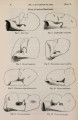
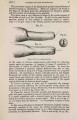
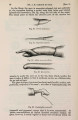
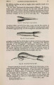
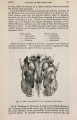
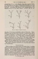
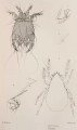
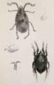
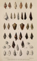
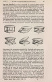
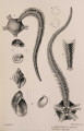
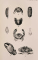
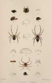
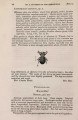
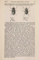
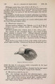
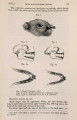
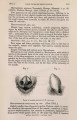
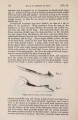
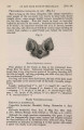
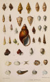
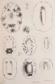
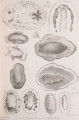
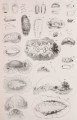
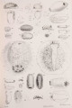
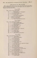
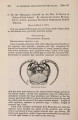
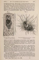
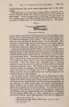
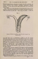
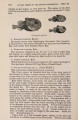
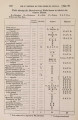
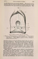
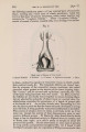
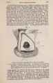
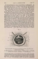
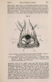
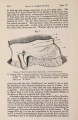
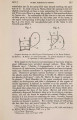
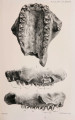
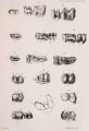
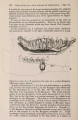
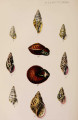
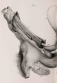
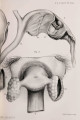
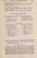
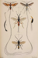
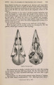
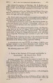
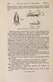
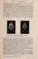
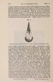
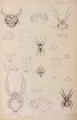
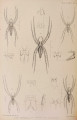
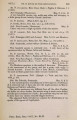
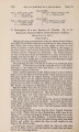
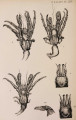
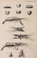
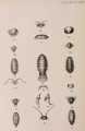
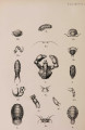
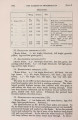
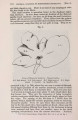
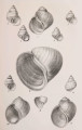
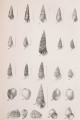
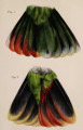
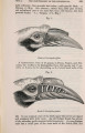
Show 678 ON CRUSTACEA, CHIEFLY FROM SOUTH AMERICA. [June 19, margin inflexed, appearing nearly straight in a dorsal view ; posterior margin with a small, median, rounded lobe received into a corresponding emargination in the anterior margin of the first segment of the body. First segment of the body longer than the succeeding. Last segment of the tail longer than broad, narrowed to its extremity, which is subacute. Coxse transverse and obtuse at their posterior extremity. Legs short, with the thigh-joints not dilated. Uropoda very slender; outer ramus very long, more than twice the length of the inner. Length 1 inch. Hab. Mauritius (R. Templeton). (Coll. Brit. Mus.) This species is at once distinguished by the form of the rami of the uropoda. The habitat, which is given as " Indian Oceau," in the ' List of Crustacea,' 1. c., is marked "Mauritius" on the label attached to the specimen. EXPLANATION OF THB PLATES. PLATE LXVI. Fig. 1. Clibanarius cayennensis, p. 657, natural size. 2. carnescens, p. 658, natural size. 3. Carapace, frontal region, eyes and antennse of Clibanarius speciosus, p. 658, magnified about twice the natural size. 3 a. Hand of the same, magnified twice natural size. 4. Clibanarius lordi, p. 658, natural size. 4 a. Carapace, frontal region, eyes and antennaB of the same, magnified twice the natural size. PLATE LXVII. Fig. 1. Palcemon jelskii, p. 661, lateral view, magnified about twice the natural size. 1 a. Carapace, frontal and antennal region of the same, dorsal view, magnified twice the natural size. 1 b. Terminal segment and uropoda, magnified. 2. Euryrhynchus wrzesniowskii, p. 662, lateral view, magnified four times the natural size. 2 a. Carapace, frontal and antennal region of the same, magnified four times the natural size. 2 b. Terminal segment and uropoda, magnified. 3. Armadillidium cmlatum, p. 665, dorsal view, magnified twice the natural size. 3 a. Dorsal view of head. 3 b. Dorsal view of tail-segments of the same, further magnified. 4. Cubaris affinis, p. 666, lateral view, magnified twice the natural size. 4 a. Dorsal view of head. 4 b. Dorsal view of tail-segments of the same, further magnified. PLATE LXVIII. Fig. 1. Cubaris gigas, p. 666, lateral view, natural size. 1 a. Head viewed from above. 1 b. Head viewed from below, showing position of the antennse. 1 c. Segments of the tail, dorsal view, all further magnified. 2. Porcellio cayennensis, p. 667, dorsal view, magnified twice the natural size. 2 a. Front view of head. 2 b. Segments of the tail, dorsal view, further magnified. 3. Porcellio jelskii, p. 668, dorsal view, magnified twice the natural size. |