| OCR Text |
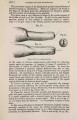
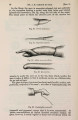
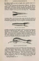
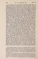
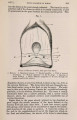
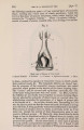
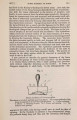
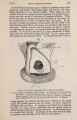
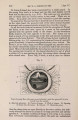
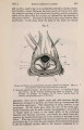
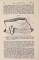
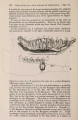
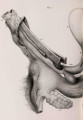
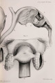
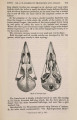
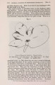
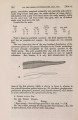
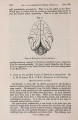
Show 1877.] ANATOMY OF HYiENA CROCUTA. 375 these structures. The two spongy bodies of opposite sides are quite distinct from one another, and do not communicate except through the vein just described ; nor do they take any part in the formation of the glans clitoridis, that body be'ing formed entirely by the corpora cavernosa and the walls of the urino-genital canal. It will also be observed that the spongy bodies are placed altogether above that canal, and that consequently the latter is not surrounded by the spongy structure in any part of its course. An artery of considerable size enters the bulbous extremity of each spongy body. Two muscles are met with in the dissection of the clitoris. The one is the erector clitoridis, which is of large size and covers the crus, as well as the bulb of the corpus spongiosum, in the usual manner. The other is the retractor clitoridis. The origin of this muscle I could not determine, as the parts had been detached from the bones ; but in all probability it arose either from the ischium or pubis. The muscles of opposite sides pass forward in contact with one another, and form two cord-like bands which lie along the lower borders of the corresponding cavernous bodies ; and each is inserted by means of an aponeurotic structure into the corresponding corpus cavernosum immediately behind the glans. These muscles are doubtless retractors of the glans within the prepuce. A large artery, vein, and nerve run along the outer side of each cavernous body, and can be traced as far as the base of the glans. There is no bone in the clitoris. If, now, we compare the description above given of these parts in Hycena crocuta with that of the corresponding organs in other species, we find, with regard to the anal-gland pouch, that its presence has been proved by Daubenton1 in the Hycena striata, by Murie2 in the Hycena brunnea and crocuta, and by Prof. Flower3 in the allied genus Proteles. With regard to H. crocuta, its presence has been denied by Busk4; but Dr. Murie's investigations, along with my own, now leave, I think, no doubt regarding this point. Anal glands are present in all of these animals, but differ somewhat in respect of number in different species. In H. striata Daubenton describes and figures two on each side of the anal-gland pouch. The anterior of these corresponds most closely with that which is present in H. crocuta, the posterior having no representative in the latter species. He further describes in H. striata a quantity of isolated follicles which have almost the same arrangement as the transverse belt of glandules which I have described as opening into the anal-gland pouch in H. crocuta. Dr. Murie describes in H. brunnea three anal glands on each side of the rectum, but makes no mention of isolated glandules in relation to the fundus of the anal pouch. Lastly, in Proteles two anal glands of large size are described by Prof. Flower, in addition to a central glandular mass covering the bottom of the anal pouch. The large glands correspond almost exactly in shape with those I have described in H. crocuta ; and although 1 am in doubt, from his description, of the exact appearance of the central glandular mass, it appears to correspond closely to the belt of 1 Buffon's Hist, Nat. torn. ix. 2 Trans. Zool. Soc. vii. p. 503. 3 Proc. Zool. Soc. 1869, p. 493. ' Quoted by Dr. Murie in his paper. |