| OCR Text |
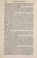
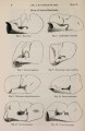
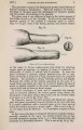
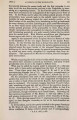
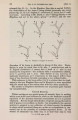
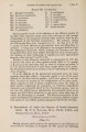
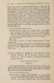
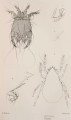
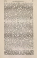
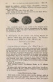
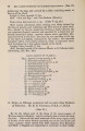
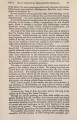
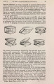
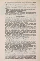
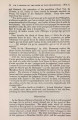
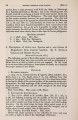
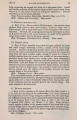
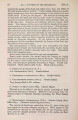
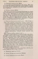
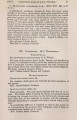
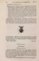
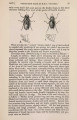
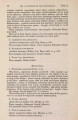
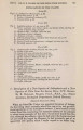
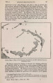
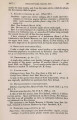
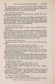
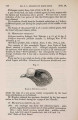
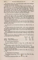
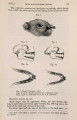
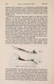
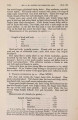
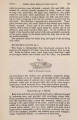
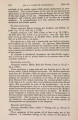
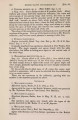
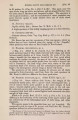
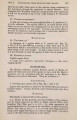
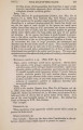
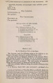
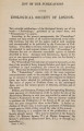
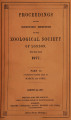
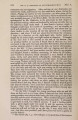
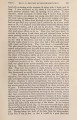
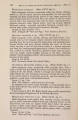
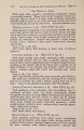
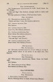
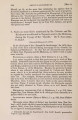
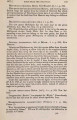
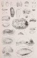
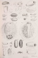
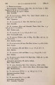
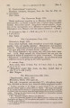
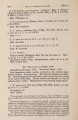
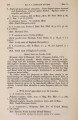
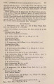
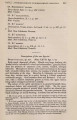
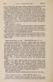
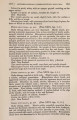
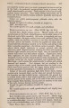
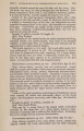
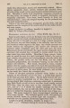
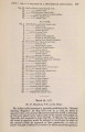
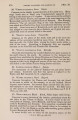
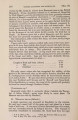
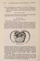
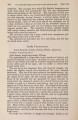
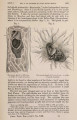
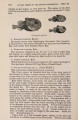
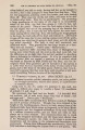
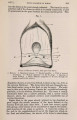
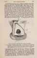
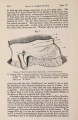
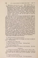
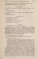
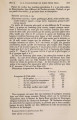
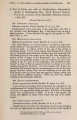
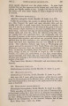
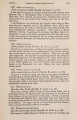
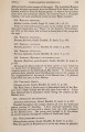
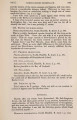
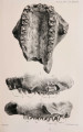
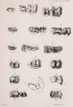
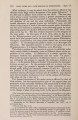
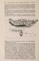
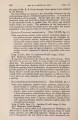
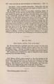
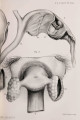
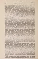
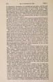
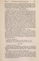
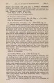
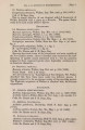
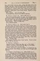
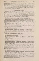
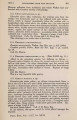
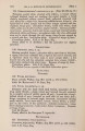
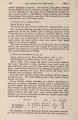
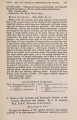
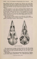
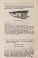
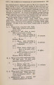
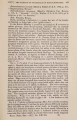
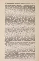
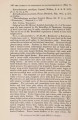
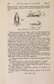
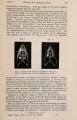
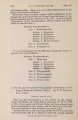
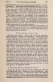
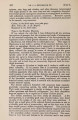
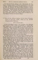
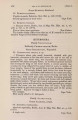
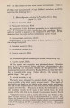
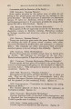
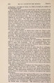
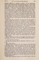
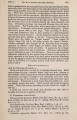
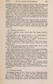
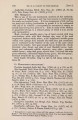
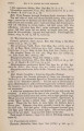
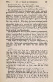
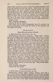
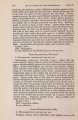
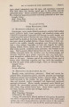
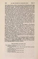
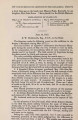
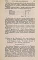
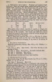
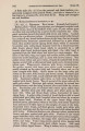
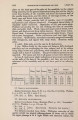
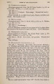
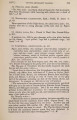
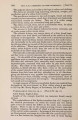
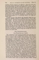
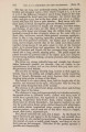
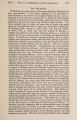
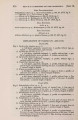
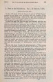
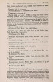
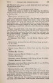
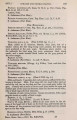
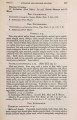
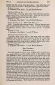
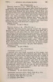
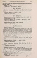
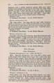
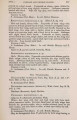
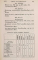
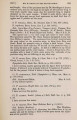
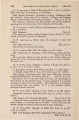
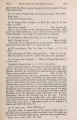
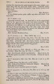
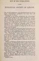
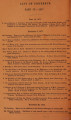
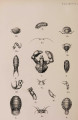
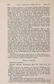
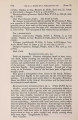
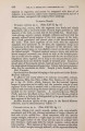
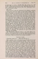
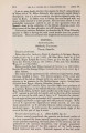
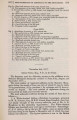
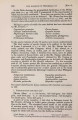
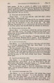
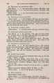
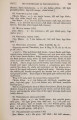
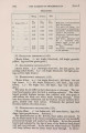
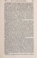
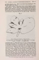
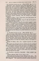
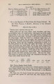
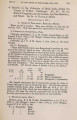
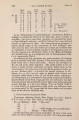
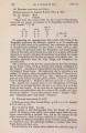
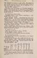
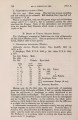
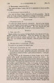
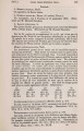
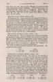
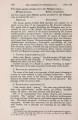
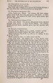
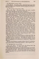
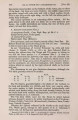
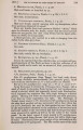
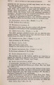
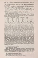
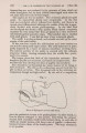
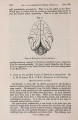
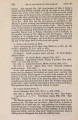
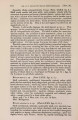
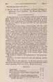
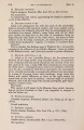
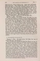
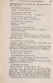
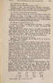
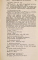
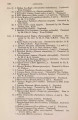
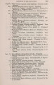
Show 310 MR. W. A. FORBES ON THE [Apr. 17, the following constituent parts :-(1) an external layer of connective tissue, covering (2) a thick layer of elastic membrane; inside this is (3) a thin layer of mucous membrane, which unites together and connects the "lymphatic follicles." These "lymphatic follicles consist of masses of minute rounded cells, on an average 0*04 millim. Fig. 2. a ---c • 1 ^ - d Back view of Cloaca of Uria troile. a. Bursa Fabricii. b. Oviduct, c, c. Ureters, d. Sphincter muscles, e. Cssca. in diam., enclosed in capsules of connective tissue, in which ramify their nutrient vessels &c. The lymphatic follicles are bound together by processes of the connective mucous membrane into raised processes, which project on the interior of the bursa, forming ridge-like " crests," and are covered with epithelium internally, the cells of the latter being lanceolate with oval nuclei. In Rhea, however, the follicles are not closely bound together in masses forming ridges, but are attached by peduncles of elastic tissue to a central stem, the whole having somewhat the appearance of a bunch of grapes with a few berries on it. As we have already seen, Tannenherg in 1789 was the first to point out that the bursa was more developed in young than old birds, it being gradually reduced and obliterated in the latter. This process of atrophy seems to obtain in all birds, so far as I can make out, though the periods of final disappearance seem to vary much in different groups. M. Martin St.-Ange found that the bursa began to lose its functional activity in Pigeons after six months, and in Fowls after eight; as a rule it seems to atrophy at about the period of full growth. On the other hand, in some cases it persists for long periods, and probably throughout life ; for I found it well developed and quite open in a specimen of Platycercus icterotis that |