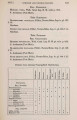
| OCR Text |
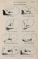
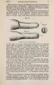
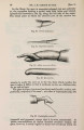
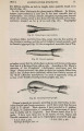
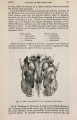
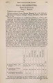
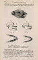
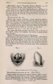
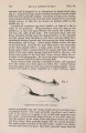
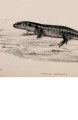
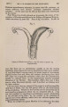
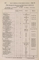
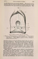
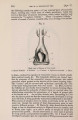
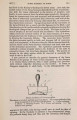
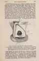
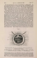
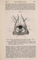
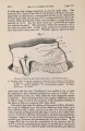
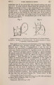
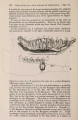
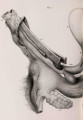
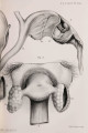
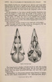
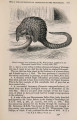
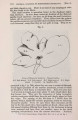
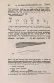
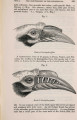
Show 372 DR. M.WATSON ON T H E [May J, the size of a small pea, and each provided with a single duct which perforates the fundus of the pouch by means of one of the little apertures previously described. Bladder and Urethra.--The bladder is small in size, and regularly pyriform. It passes insensibly into the urethra ; so that it is difficult to say where the former ends and the latter begins. It is, with the exception of the base, entirely covered with peritoneum, which forms three ligaments to the viscus. Two of these pass upward to connect it with the uterine cornua, and one downwards to attach it to the anterior abdominal wall. The ureters are provided with thick muscular walls, and enter ihe bladder close to the orifice of the urethra. This tube measures only 1^ inch in length, and passes backwards, resting against the corpus uteri to open into the urogenital canal, its orifice being separated from the os uteri by a single semilunar fold of mucous membrane. Ovaries.-Each of these bodies is of an oval flattened form, having its long axis placed transversely, and its anterior and posterior surfaces somewhat flattened. Each measures rather more than half an inch in length and about a fourth of an inch in thickness. The ovary is retained in position by two stout ligaments, one of which is attached to the inner, the other to its outer extremity. The former attaches the ovary to the outer extremity of the corresponding uterine horn, and measures half an inch in length. By means of the latter, which measures 5 inches in length, the ovary is fixed to the posterior surface of the diaphragm. Each of these ligaments consists of a fibrous cord, and each is covered by peritoneum. The ovary is placed in a peritoneal pouch of size sufficient to contain a large walnut. The opening of this pouch is considerably narrowed, and in the natural position of the parts is directed upwards (towards the spine of the animal) so that ova, as they pass from the surface of the ovary must almost necessarily be caught by it. This pouch is formed by a reduplication of the broad ligament of the uterus, and appears to be, so to speak, suspended from the uterine and diaphragmatic ligaments of the ovary, hanging vertically below that organ, which is placed just within the entrance to the sac. The posterior free margin of the pouch is formed by the ovary together with its uterine and diaphragmatic ligaments, whilst its anterior margin is formed by the free edge of the peritoneal fold and, to a less extent, by the fimbriated extremity of the Fallopian tube. Fallopian Tube.-In order to be explicit, I must here state that by the term Fallopian tube I understand so much of the oviduct as extends from its fimbriated extremity to the point of attachment to it of the uterine ligament of the ovary. That portion of the oviduct which extends from the attachment of this ligament to its junction with its fellow of the opposite side, 1 regard as constituting the horn of the uterus. The Fallopian tube, as thus defined, measures between 2\ and 3 inches in length, and describes a U-shaped flexure from the one extremity to the other, the concavity of the U in the natural position of the parts being directed upward. The free extremity of the Fallopian tube lies in contact with the anterior free margin of |