| OCR Text |
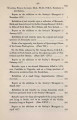
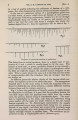
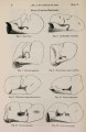
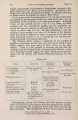
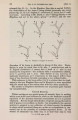
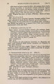
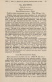
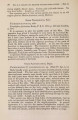
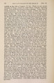
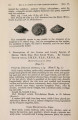
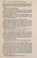
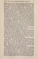
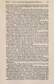
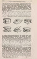
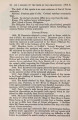
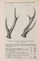
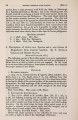
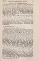
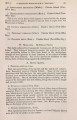
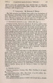
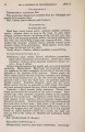
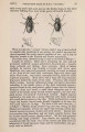
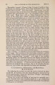
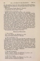
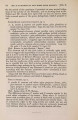
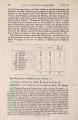
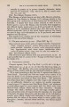
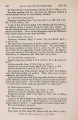
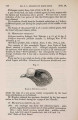
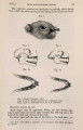
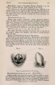
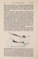
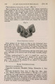
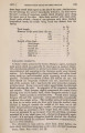
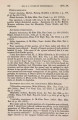
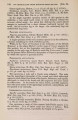
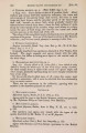
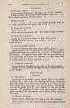
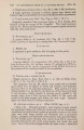
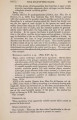
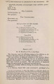
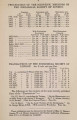
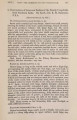
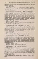
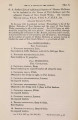
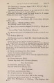
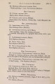
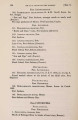
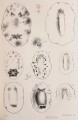
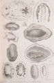
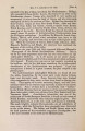
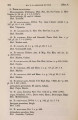
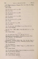
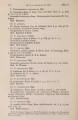
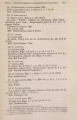
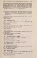
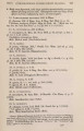
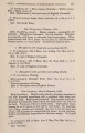
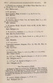
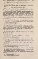
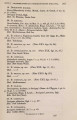
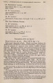
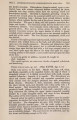
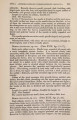
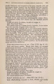
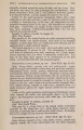
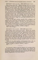
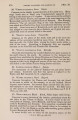
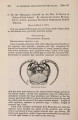
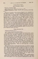
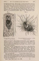
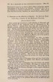
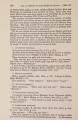
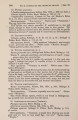
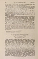
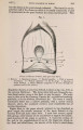
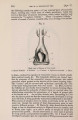
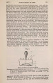
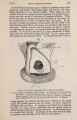
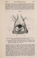
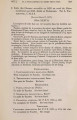
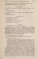
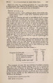
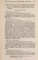
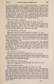
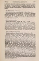
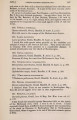
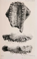
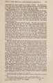
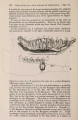
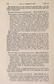
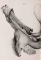
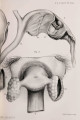
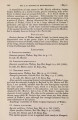
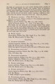
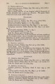
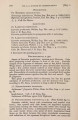
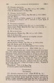
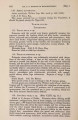
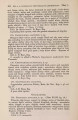
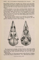
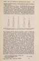
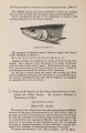
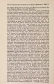
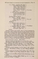
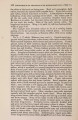
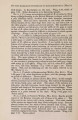
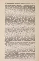
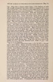
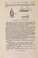
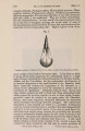
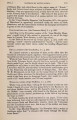
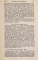
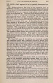
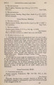
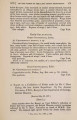
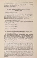
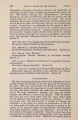
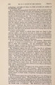
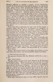
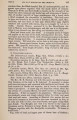
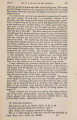
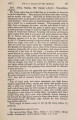
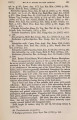
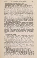
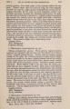
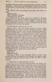
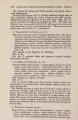
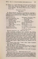
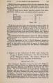
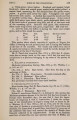
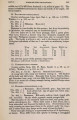
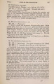
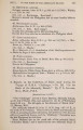
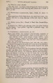
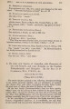
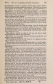
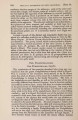
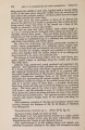
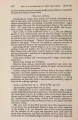
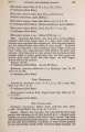
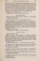
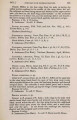
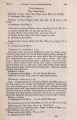
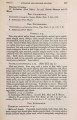
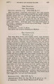
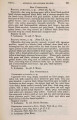
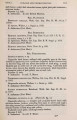
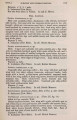
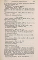
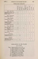
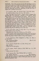
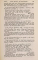
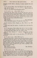
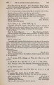
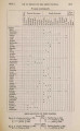
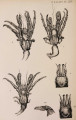
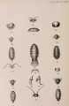
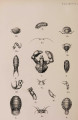
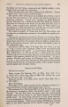
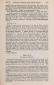
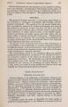
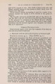
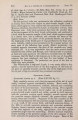
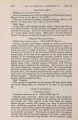
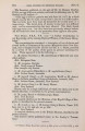
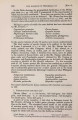
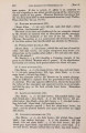
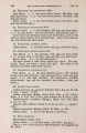
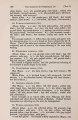
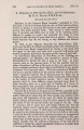
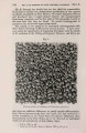
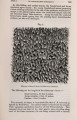
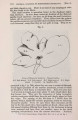
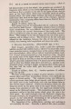
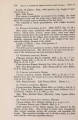
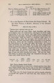
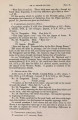
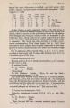
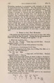
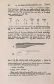
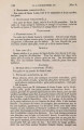
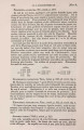
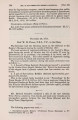
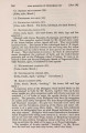
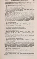
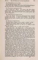
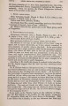
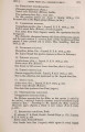
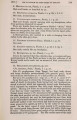
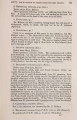
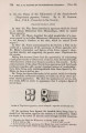
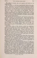
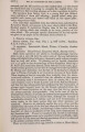
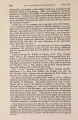
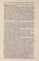
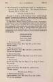
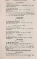
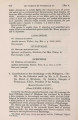
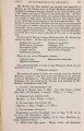
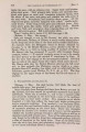
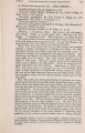
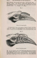
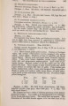
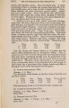
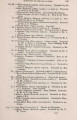
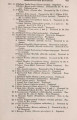
Show 1877.] BURSA FABRICII IN BIRDS. 315 and in situ,-and in fig. 6, in an Ostrich (Struthio comelus, immature female)-where the bursa has been nearly all removed to show the posterior opening of the cloaca into its cavity, and the communication of the latter with the exterior, as indicated by the direction of the pointer (D D'). The same is the case in the young Nandou Rhea americana). In all these birds the walls of the bursa are thickly Fig. 6. Cloaca and Bursa of young Ostrich (female), viewed from behind. Most of the posterior wall of the bursa has been removed. A. External sphincter muscle. B. Cut surface of bursa. C. Opening of cloaca into bursa. D, D'. Pointer passing from bursa to exterior. E, E. Ureters. F. Oviduct. G. Clitoris. H, H'. Pores; beneath them the smooth, non-glandular part of the bursa. glandular; there are no regular crests and sulci, however, but the glands are arranged in patches, the whole having a honeycomb-like or dendritic appearance. This disposition of parts, however, is not permanent. As the birds grow older, the size of the bursa gradually diminishes and its walls become less glandular; its mouth is no longer equal in extent to the whole width of the outermost chamber, but becomes narrowed; and finally the whole bursa disappears, its remains becoming lost in the muscles of the back of the cloaca. This state of atrophy of the bursa is represented in Casuarius picticollis in fig. 7 (p. 316), the only remains of its existence being seen in the few irregular circular folds on the mucous membrane at A. |