| OCR Text |
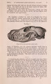
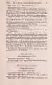
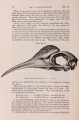
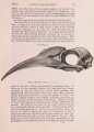
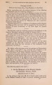
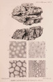
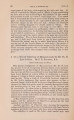
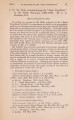
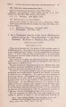
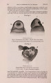
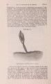
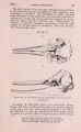
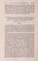
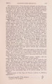
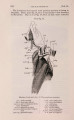
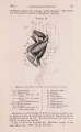
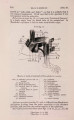
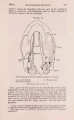
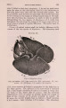
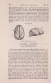
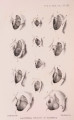
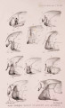
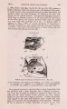
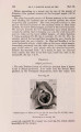
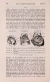
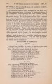
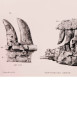
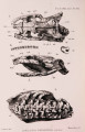
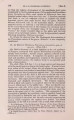
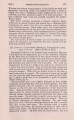
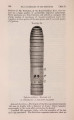
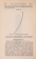
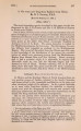
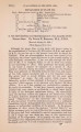
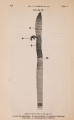
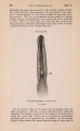
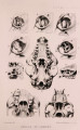
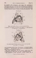
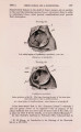
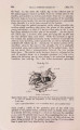
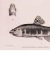
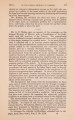
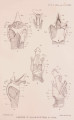
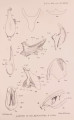
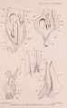
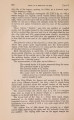
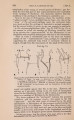
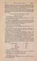
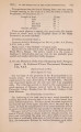
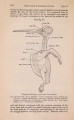
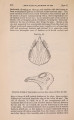
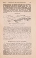
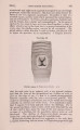
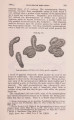
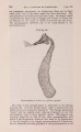
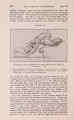
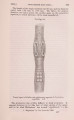
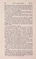
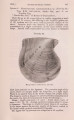
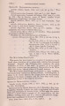
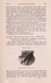
Show 266 DR. 0. I. FORSYTH MAJOR O N [Mar. 19, the skull. In the orbit (PI. XXII. fig. 1) tbe inflated part of the palatal appears situated medially from the orbital plate of the maxilla, and medially as well as posteriorly from the os planum ; the latter helps to form the posterior prolongation of the maxillary sinus and covers besides the anterior prolongation of a sphenoidal sinus, laterally it is in its turn covered by the frontal. In this stage the palatal pneumatic cavity rather resembles the swollen orbital maxillary plate above the germs of the molars of young individuals; so that, if it has ever been seen at all, it m ay have been mistaken for that part of the maxilla. As the figures (PJ. X X I I . figs. 1, 2) show, both parts are lying side by side and are very distinct from each other. With the increase in age of the animal, the palatal pneumatic cavity continues to pervade the orbit in every direction, antero-posteriorly as well as laterally and medially (PI. X X I I . fig. 3). Anteriorly, the hinder portion of the maxillary sinus is the loser in the struggle, for it is gradually encroached upon and pushed forward by the palatal sinus; but I am not aware that a communication between the two cavities takes place. The palatal cavity becomes, however, enlarged at the cost of another sinus; in a youngish specimen (m.3 not yet in place) the following can be seen owing to its somewhat damaged condition (see text-fig. 70):-the before-mentioned sphenoidal Text-fig. 70. Lemur rubriventer. Right orbital region. The bones forming the floor have been partially removed, in order to exhibit the disposition of the underlying sinuses. About % nat. size. s.sph.- sphenoidal 6inus; s.mx. = maxillary sinus; spa. = palatal sinus. sinus (s.sph.), which iu the posterior part of the orbit runs parallel to the palatal cavity, medially from it, finally turns round in a lateral direction, ending in a cul-de-sac between the maxillary (s.mx.) and the palatal sinus (s.pa.), thus separating the two. As in later stages I have found no more trace of this cul-de-sac, its place being occupied by the palatal sinus (text -fig. 70), it is evident that it must have been absorbed by the latter. |