| OCR Text |
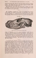
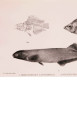
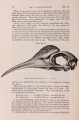
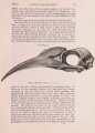
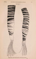
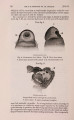
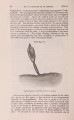
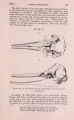
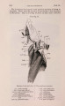
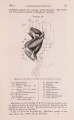
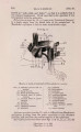
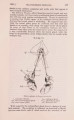
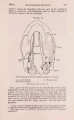
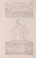
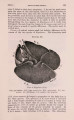
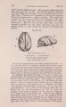
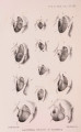
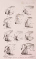
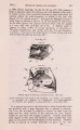
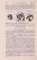
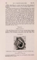
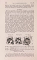
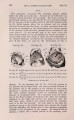
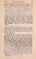
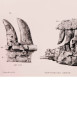
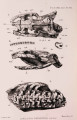
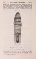
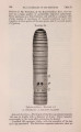
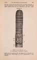
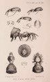
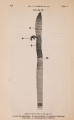
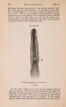
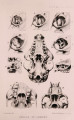
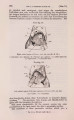
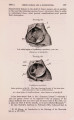
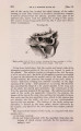
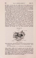
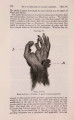
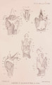
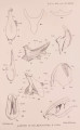
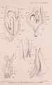
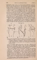
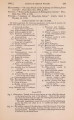
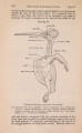
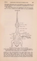
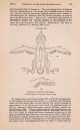
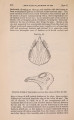
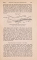
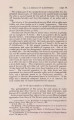
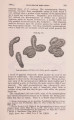
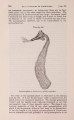
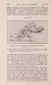
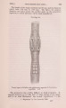
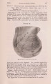
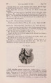
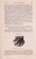
Show 1901.] LARYNX OF CERTAIN WHALES. 293 but also to the outer surface. Only a short piece of the muscle remained in connection with the larynx, but the anterior direction of the fibres and the position of its attachment render it probable that it is the muscle of this name. 2. Just below this is the sterno-thyroid muscle (S.t.), the fibres of which pass obliquely backwards and downwards towards the position occupied by the sternum. 3. Separated from this muscle by a sheet of fibrous tissue is a third large muscle (x) whose identification is uncertain. The fibres are directed antero-ventrally, i. e. downwards and somewhat forwards, though the inclination is but slight, and they pass nearly directly ventralwards. This mass of muscle is attached over nearly the whole of the lower half of the thyroid plate between the " bay " and the ventral margin, which, however, it does not reach. As the larynx had been cut away from the neighbouring organs, and indeed cut across near the lower end, I am unable to identify the muscle : perhaps it is an accessory sterno-thyroid. 4. The dorsal edge of the posterior cornua and of the thyroid plates also serves for the attachment of muscles, probably the stylo-pharyngeal and the basio-thyro-hyoid (cf. Macalister, 1867). 5. Crico-lhyroid muscle.-This is very small in Cogia and invisible from without, as it is entirely concealed, partly by the posterior cornu and partly by a fan-shaped tendon that passes from its ventral edge across the " bay " to the thyroid plate. But when this tendon is removed, a small muscle is exhibited (PI. X X V . fig. 4, PI. X X V I . fig. 6 a, C.t.). In its diminutive size it contrasts very notably with the homologous muscle in Balcenoptera, and indicates a very feeble mobility of tbe thyroid cartilage upon the cricoid- In some Odontocetes, e. g. Globicephalus melas, according to Murie (1867), this muscle is of " considerable size," while Macalister mentions that in G. svineval the crico-thyroid is attached " to the posterior edge of the thyroid cartilage," and makes no mention of its attachment to the cornu. 5. The crico-arytenoid muscle is here represented by a posterior and lateral division (the latter being absent in Mystacocetes). The posterior muscle (PI. X X V . fig. 5, Car.) is a large quadrate mass arising from the dorsal face of the cricoid and passing forwards to the arytenoid, to the "processus muscularis" to which it is attached. . The lateral division (PI. XXVII. fig. 18) arises from the side of the cricoid, ventral of the thyroid facet, and some of its fibres arise from the horn of the thyroid (as Murie states is also the case in Globiocephalus), and indicating the close relation of this muscle to the thyro-arytenoid. 6. The transverse arytenoid muscle is a thin sheet having the usual relations, and forming the dorsal wall of the " aryteno-epiglottidean tube." 7. The aryteno-epiglottid muscle (PI. XXVII. fig. 18, A.ep.) is comparatively small, and connects the lower regions only of the two cartilages. 8. Above this is a much stouter muscle, the thyro-epiglottid(T.ep.), which arises from the inner surface of the thyroid near its ventral PROC. ZOOL. Soc-1901, VOL. I. No. XX." 20 |