| OCR Text |
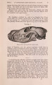
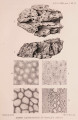
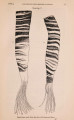
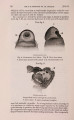
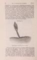
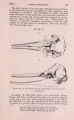
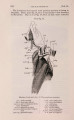
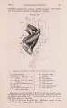
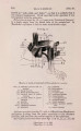
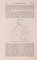
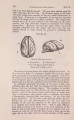
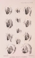
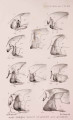
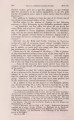
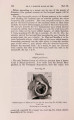
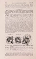
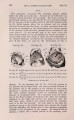
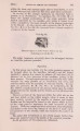
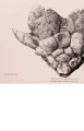
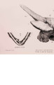
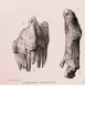
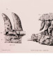
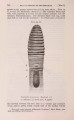
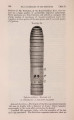
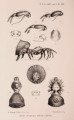
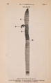
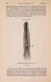
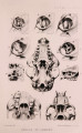
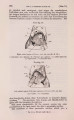
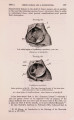
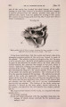
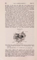
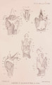
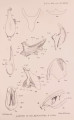
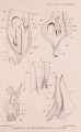
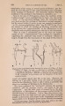
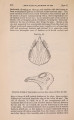
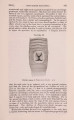
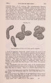
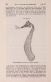
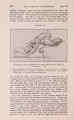
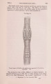
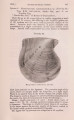
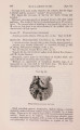
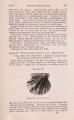
Show 290 PROF. W. B. BENHAM ON THE [Apr. 2, one side of the larynx, meeting its fellow at a distinct angle, and so forming a ridge. The body is irregularly rectaugular (PL X X V I . fig. 6), with a nearly straight but slightly curved ventral border having a thin edge, a curved aaterior border presenting a recurved and thickened edge, which passes dorsally with the posterior cornu ; the posterior border of each ala is oblique but straight, while the dorsal border is curved, and passes forwards to join the root of the posterior cornu. These various " borders" pass into one another at rounded angles, but the angle formed by the ventral and posterior borders is better marked than the rest, and it is at this angle that the two alae-the right and left-approximate most closely; nevertheless they only just meet, and this when the apparatus is at rest. It is here that the lower end of the epiglottis rests, as will be seen later. The posterior cornu of the thyroid is a short, flat, narrow plate, whose base passes quite imperceptibly into the dorso-anterior region of tbe body, but between the cornu and the dorsal border of the plate there is a well-marked " bay." Each thyroid plate is nearly flat; it is only feebly convex in a dorso-ventral direction (a convexity which is slightly exaggerated in the figure of the ventral view); the edge is thin, except along the anterior border, which is thick and everted, and probably represents the " anterior cornu." The measurements of this plate are as follows :- The ventral border is 3 inches, measured along the curve. The posterior border is 2\ inches. The anterior border is 1 | inches. The dorsal (behind the cornu) is 2 inches. The outer curve of the posterior cornu is 4^ inches, while its inner (i. e. ventral) margin is about H inches, and its breadth f inch. In the Pilot Whale the figures and account given by Murie show a very different thyroid ; the body, which is single, being transversely extended across the ventral surface of the larynx, while the posterior cornua are much longer, leaving a deep wide bay on each side between themselves aud the body. The cricoid cartilage (PI. XXATI. figs. 8, 9, 10) is a complete ring, and, as usual, is of greater height (i. e. antero-posterior length) on its dorsal half than on its ventral. The dorsal half of the riug is a broad thick band, deeply excavated on its binder margin, while its anterior margin is irregularly convex ; when viewed from this aspect, then, it has the appearance of an inverted V with a very open angle (about 90°) and thick limbs. The median line of this dorsal face projects as a slight convex ridge, separating the right and left muscular fossae from one another. The sloping sides bear on the upper margin the |