| OCR Text |
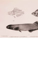
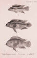
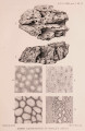
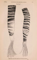
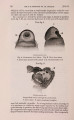
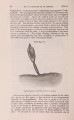
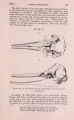
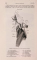
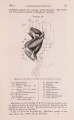
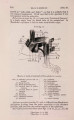
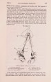
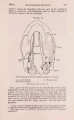
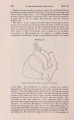
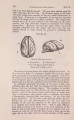
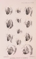
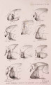
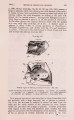
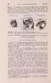
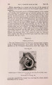
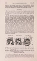
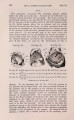
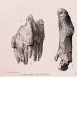
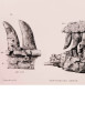
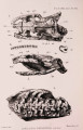
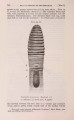
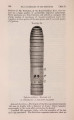
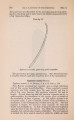
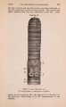
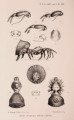
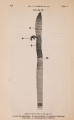
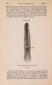
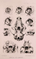
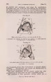
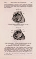
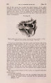
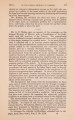
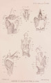
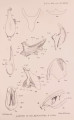
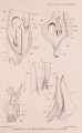
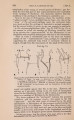
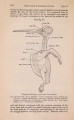
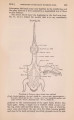
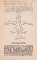
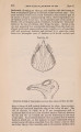
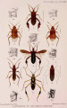
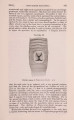
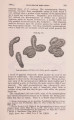
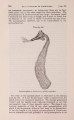
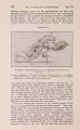
Show 300 ON THE LARYNX OP CERTAIN WHALES. [Apr. 2, Fig. 2. Dorsal view of the larynx of the same. Some of the muscles have been removed from the right side. Note the continuity of the upper tracheal rings with tbe cricoid cartilage. Fig 3. View of the right side of the larynx of the same. Fig. 4. Cogia. Ventral view of the larynx, the extrinsic muscles of the left side having been removed. The thyroid cartilage is seen to consist of separate right and left abe or plates, which have been somewhat forced apart in order to show the subepiglottid cartilage. On the left side, the tendon (W. on tbe right) had been cut away so that the small crico-tbyroid muscle is partially exposed. Fig. 5. Dorsal view of the larynx of the same. PLATE XXVI. Fig. 0. The larynx of Cogia from the right side. Fig. 6 a. Aperture of the same, showing the small crico-thyroid muscle, the outline of which is dotted, where it is hidden by tbe thyroid cornu. Fig. 7. Balanoptera. The cricoid cartilage from the ventral surface. Fig. 8. Cogia. The cricoid cartilage (ventral view). Fig. 9. Tbe same, dorsal view. Fig. 10. The same, side view. r. ridge, along which the lateral crico-arytenoid is inserted. Fig. 11. Balcenoptera : the epiglottid cartilage from within (i. e. dorsal surface). a, the upper and b, the lower region. Fig. 12. The same, side view, in situ (cf. fig. 17): a, b, transverse sections of tbe epiglottid cartilage. Fig. 13. Cogia. The epiglottis from tbe side. PLATE XXVII. Fig. 14. The epiglottis of Cogia from within: a, b, transverse sections at the levels similarly marked. Fig. 15. Balanoptera. Tbe right arytenoid cartilage from without. Fig. 16. Cogia. The right arytenoid cartilage from without. Fig. 17. Balcenoptera. The aryteno-epiglottidean apparatus, seen after removal. of the right half of the thyroid cartilage. Fig. 18. Cogia. The aryteno-epiglottidean tube, from the right side, after removal of tbe right ala of the thyroid. The cricoid cartilage had been cut and the epiglottis has sunk downwards slightly. Fig. 19. Cogia. Tbe inner face of the left thyroid plate, showing muscle attachments. Fig. 20. Cogia. The entrance to the larynx, at the apex of the aryteno-epiglottidean tube. Fig. 21. Balcenoptera. The right side of the pharynx has been cut through, and carefully lifted without disturbing the relations of the epiglottis aud palate. P L A T E XXVIII. Fig. 22. Balanoptera. The same as fig. 21, after the depression of the floor of the pharynx, so that the entrance to the larynx is displayed. Fig. 23. Balanoptera. The sublaryngeal pouch has been opened by a longitudinal incision ; tbe right arytenoid has been turned upwards so as to open out the groove between them and expose the glottis, the opening between the pouch and the larynx. This aperture (0.) is placed between the posterior processes of the arytenoid cartilages. Fig. 24. Balcenoptera. Outline of larynx, supposed to be transparent, so as to exhibit tbe extent and relations of tbe sublaryngeal pouch and the cavity of the larynx : the contained cavities are shaded. Fig. 25. Cogia. View of the interior of the aryteno-epiglottid tube, as seen when it has been opened by an incision separating the right arytenoid from the epiglottid. It is seen that there is no median sac at the base of the epiglottis. Fig. 26. Ziphius, sp. A median longitudinal section through the larynx. t, tubular outgrowths from the lateral pouches ; s, membranous septum separating the right and left pouches. |