| OCR Text |
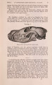
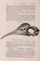
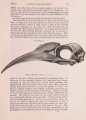
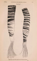
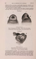
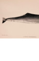
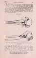
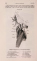
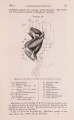
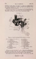
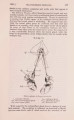
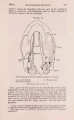
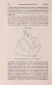
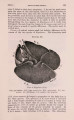
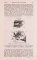
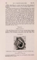
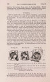
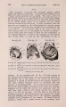
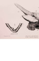
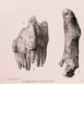
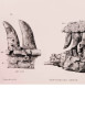
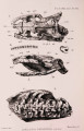
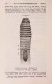
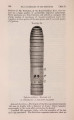
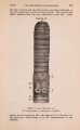
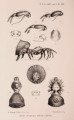
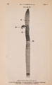
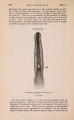
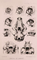
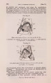
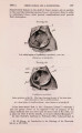
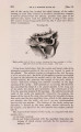
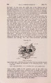
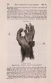
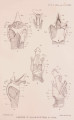
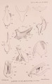
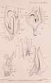
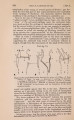
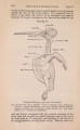
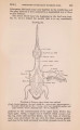
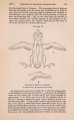
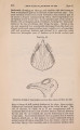
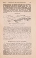
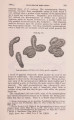
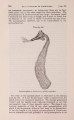
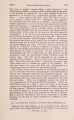
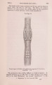
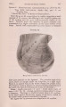
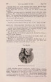
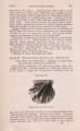
Show 1901.] LARYNX OP CERTAIN WHALES. 289 is, of course, formed by the tip of the epiglottis, also thick and rounded. This aperture is, then, perfectly well defined; it measures two inches by one inch, but it is rather wider at the ventral than at the dorsal (arytenoid) end. As seen from the side, this tube is somewhat peniform, Ihe free end terminating in the thick lips just referred to, the arytenoids projecting beyond the epiglottis. I have seen no trace or indication of a sublaryngeal pouch in Cogia, such as has been described by Murie (1871) for Bisso's Grampus, by Watson and Young (1879) for Beluga, and by Sir W m . Turner (1886) for Mesoplodon. Murie writes (p. 127), near the base of the epiglottis there is " a median orifice leading into a moderate-sized pouch, which fills in great part the angle of junction between the enlarged epiglottis and the thyroid cartilage ; " and Turner says (p. 165) that between the forks of the bifurcated epiglottis and the upper border of the thyroid cartilage there is a shallow mesial pouch, lined by mucous membrane, which freely communicates with the interior of the larynx. With these statements before me I looked carefully for this pouch in Cogia, but it is absent. There is no space or " angle" between the epiglottis and the thyroid such as Murie describes, and there seems to be actually " no room " for any such pouch. At any rate, there is none, nor is there any glandular tissue to represent it, which Murie describes and figures (p. 128) in relation to the pouch. In Cogia the lining membrane both of the arytenoids and of the epiglottis is smooth ; the median ridge on the latter forms a slight depression on each side (which is precisely what occurs, too, in Balcenoptera), aud in the lower half of these lateral grooves tbe mucous membrane is pitted ; these small pits and depressions are, however, present only on the sides, not in the middle line as Murie describes for Bisso's Grampus. Nor does he mention any pouch in Gl. melas (1867), nor do I find one in Ziphius (see below). The Cartilages. The tracheal rings are here complete, and the upper ones present certain irregularities that will be better understood by reference to the figures than by a description. About one inch below the larynx the trachea gives off on the right side a bronchus-the third bronchus-as in most other Odontocetes. The cartilages, as will be seen by a glance at the figures, differ very considerably in form and proportions from the correspondiug parts in the Borqual. The thyroid cartilage (PL X X V . fig. 4, T.) is represented by two separate pieces, a right and a left, which meet ventrally. These two halves may be termed for convenience the thyroid plates or alse. Each thyroid plate presents a " body " and cornu, and forms |