| OCR Text |
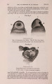
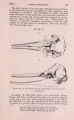
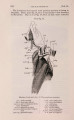
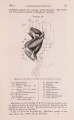
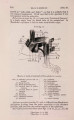
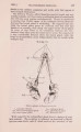
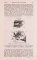
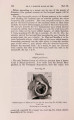
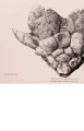
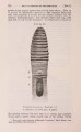
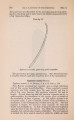
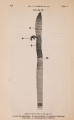
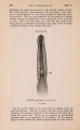
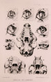
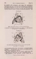
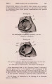
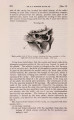
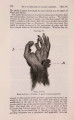
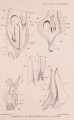
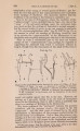
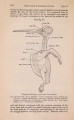
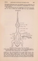
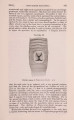
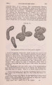
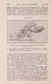
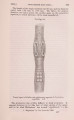
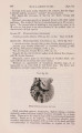
Show 1001.]] SUPRAORBITAL CANAL OE CHIMERA. 185 Teleosts and one or two Elasmobranchs the nerve to the anterior organ of the main trunk-canal emerges from the brain in connection with the glossopharyngeal; and iu Chimcera two organs in the middle of the supraorbital canal are innervated by twigs from the Ramus ophthalmicus profundus of the Vth cranial nerve-apparently the only genuine case of connection between the nerves of the lateral line and the trigeminus. This anomaly in the innervation of the supraorbital canal in Chimcera was discovered by Cole1, and evidently caused him considerable perplexity, for he does his best to minimise the awkwardness of the fact and calls to his aid a suggestion thrown out by Pollard to the following effect:-"I should prefer to say that some nerve-fibres had struck the path of the profundus but did not belong to it, just as, for instance, in Siluroids the fourth nerve accompanies the profundus, though I think everyone would hesitate to say that the fourth nerve was a branch of the profundus "2. This suggestion, ingenious as it is, cannot without further evidence be said to give us much practical help. In the following note I hope to be able to give that further evidence and to showthat Pollard was upon the right track, although the details of the connection between the superficialis and profundus fibres do not exactly conform to the picture that he evidently had in mind. During the last few months I have had occasion to dissect three heads of Chimcera monstrosa for various purposes connected with the Museum, and in all three specimens the branch of the profundus that is said by Cole to innervate two organs of the supraorbital canal was joined after leaving the orbit by two twigs from the Ramus ophthalmicus superficialis of the facial. The figure given below (text-fig. 49, p. 186) is compounded from two of the most satisfactory dissections, in one of which the connection between the nerves, and in the other their further distribution was seen to the best advantage. O n a level with the anterior border of the interorbital membrane, the Ramus ophthalmicus profundus of the trigeminal gives off a branch as described by Cole, which runs in an antero-dorsal direction towards the forehead closely applied to the perichondrium. Shortly after leaving the orbit it divides into two subsidiary branches (A and B). The branch A, after crossing the main trunk of the superficialis VII (at this point embedded in the cartilage of the skull), again divides into two smaller twigs (C & D). The twig C continues in an almost perpendicular line towards the dorsal surface of the head and is lost in the frontal clasper in the male3, and in the female upon the skin in the correspondiug position. The twig D, on the other hand, reunites at an acute 1 Trans. R. Soc. Edinburgh, xxxviii. 1897, p. 645. 2 Trans. R. Soc. Edinburgh, xxxviii. p. 638. 3 In the male specimen, I was under the impression that this nerve to the clasper was joined by a filament from the superficialis-making, in all, tbree connections between the superficialis and profundus, but the dissection was not sufficiently good to be quite sure upon the point. |