| OCR Text |
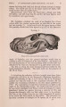
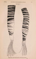
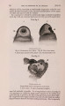
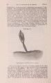
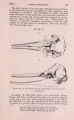
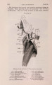
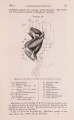
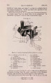
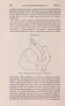
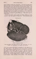
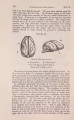
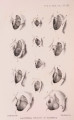
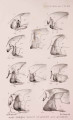
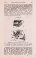
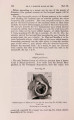
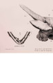
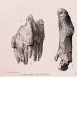
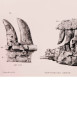
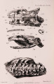
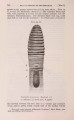
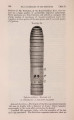
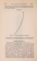
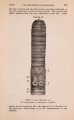
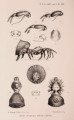
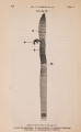
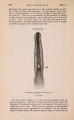
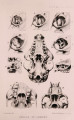
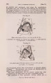
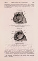
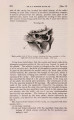
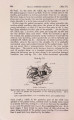
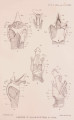
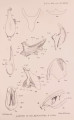
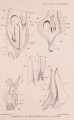
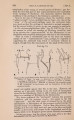
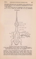
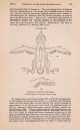
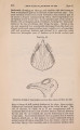
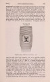
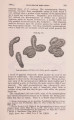
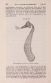
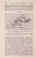
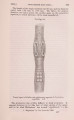
Show 292 PROF. W. B. BENHAM ON THE [Apr. 2, The region that represents the " processus muscularis" of Balcenoptera is here a slight, nearly circular, convex prominence, not at all well marked. The internal face of the entire cartilage is smooth. The length of the arytenoid is 5| inches ; its greatest breadth is 1| inches; while its upper moiety is only five-eighths of an inch across. The epiglottid cartilage (PI. X X V I . fig. 13, PI. X X V I I . fig. 14) has the usual trough-like character; it is, in contrast with that of Balcenoptera, very massive, being 6 inches in length, and its greatest breadth is 1 | inches. Seen in side view, it is club-shaped in outline ; the upper, narrower region being somewhat flattened from side to side, while the broader posterior region is much compressed ; at the junction of these two regions the hyo-epiglottid muscle is inserted. This lower region is rounded posteriorly, where it abuts against the thyroid plates. The lateral surfaces are here somewhat excavated, serving for the attachment of muscles. These surfaces meet in a relatively sharp ventral edge. The upper end of the cartilage becomes quite thin, and the extreme upper margin is recurved. Tbe posterior or internal surface is grooved ; this groove at its commencement is shallow and wide, but further down becomes deeper and narrower. Bising from the floor of the groove in the upper half is a ridge, which fades away posteriorly ; thus a transverse section near the upper region is W-shaped, while lower down it is V-shaped. The broad base of the epiglottid cartilage is capped by two small cartilages : one is patelliform, measuring f x \ inch, and is thrust between the two thyroid plates so as to be visible when the ventral margins of these are parted (PI. X X V . fig. 4 ) ; the second is smaller, oval, and nodular in form, situated dorsal of the first; it measures three-eighths of an inch long, and is closely related to the ventral edge of the right thyroid plate, connected to it by fibrous tissue. It is situated at the origin of the thyro-arytenoid muscle of the right side, aud rests against a small hard prominence on the inner surface of the ventral edge of the left thyroid plate. Each of these two subepiglottid cartilages is separated from the epiglottis by the thickened layer of fibrous tissue. It is possible that they represent the " lobular " of the 4th and 5th visceral arch, one of which persists in Echidna. I saw no representative of the process (marked c in Howes's figures) passing inwards from the base of the epiglottis towards the base of the arytenoid, to which it is connected by fibrous tissue. Muscles of the Larynx. The outer surface of each thyroid plate serves for the attachment of three muscles (PI. X X V I . fig. 6). 1. The thyro-hyoidmuscle(T.h.) is attached over the whole breadth of the anterior region of the plate, partly to the thickened edge, |