| OCR Text |
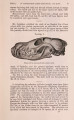
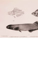
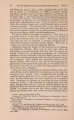
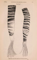
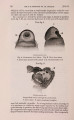
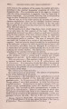
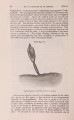
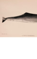
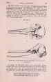
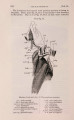
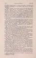
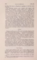
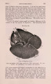
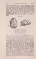
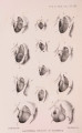
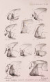
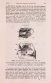
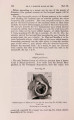
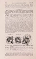
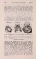
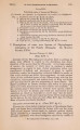
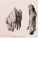
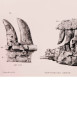
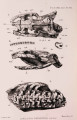
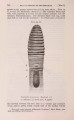
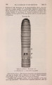
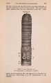
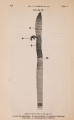
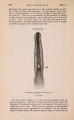
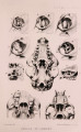
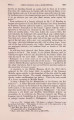
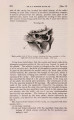
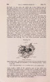
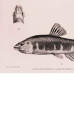
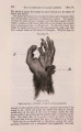
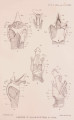
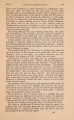
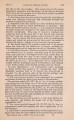
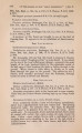
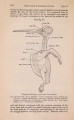
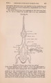
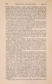
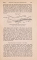
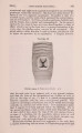
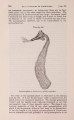
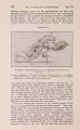
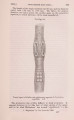
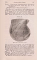
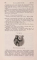
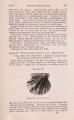
Show 1901.] LARYNX OP CERTAIN WHALES. 285 to the thyroid cartilage, and whose lower end is seen projecting through the muscles (PI. X X V . fig. 1 ) ; the upper end is in this young Borqual very thin, and the margin is reflected to support the overlying mucous membrane ; the ridge supporting the "cushion" is of short extent and does not reach the upper end. It measures 3^ inches in length; f inch deep at its base, wdiich is \ inch wide, while the upper region is f inch wide. Before passing to a consideration of the muscles connected with the cartilages, reference may be made to the form of the laryngeal cartilages in Balcena mgsticetus, which are fully and beautifully figured by Eschricht and Bernhardt (1866). The form of the cartilages is very similar in the Bight Whale to those of the Borqual, though, as would be expected, tbe proportions of the various cartilages are slightly, but not markedly, different; the only important divergences are that the posterior processes of the right and left arytenoids are united behind the entrance to the larynx, and the smaller size of the epiglottid cartilage, while the body of the thyroid is of very much greater extent than in the Borqual; nevertheless these two members of the Mystacocetes have a larynx formed on one plan, and this plan is very different from that of the Odontocetes. The Musculature of the Larynx. Drs. Carte and Macalister gave a detailed account of the various muscles of the larynx-both extrinsic and intrinsic-for _BaZceno^era,andIhavemacle no attempt here to go over this ground. I shall content myself with referring to those only that are conspicuous in this whale, and those that are of interest in contrast with the larynx of Cogia. Carte and Macalister recognize 17 muscles, intrinsic muscles, in the larynx ; most of these I have identified. 1. The crico-thyroid muscle (PI. X X V . fig. 1, C.t.) is of considerable size ; it arises from the hinder half of the latero-ventral face of the cricoid (body); the muscle-fibres pass forwards and outwards, diverging as they go, to be inserted on the inner surface of the posterior cornu of the thyroid. 2. The ventral surface of the larynx is occupied by a great bundle of muscle, longitudinally disposed in the middle line; on dissection it is found that this mass of muscle forms part of the wall of the sublaryugeal pouch, and can readily be separated into an external layer of longitudinal muscles and an inner sheet of circular fibres. Carte and Macalister describe and figure only the latter, and state that " the thick walls are almost entirely composed of circular fibres." (a) The longitudinal muscles of this sublaryngeal sac take their origin in the body of the thyroid, to which they are attached in the sides of the V-shaped notch (PI. X X V . fig. 1, T.c.) and on the inner face in the inid-line. From their point of origin the fibres spread out on both sides, forming two more or less distinctly |