| OCR Text |
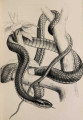
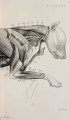
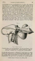
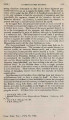
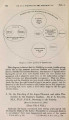
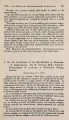
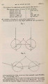
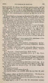
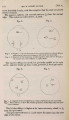
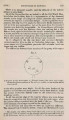
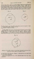
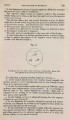
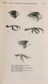
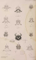
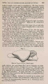
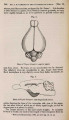
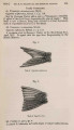
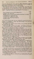
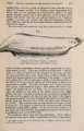
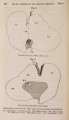
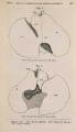
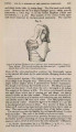
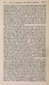
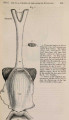
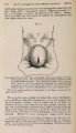
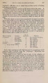
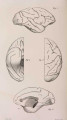
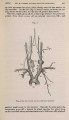
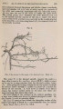
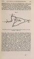
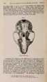
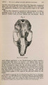
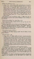
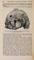
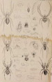
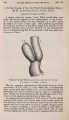
Show 466 MR. W. OTTLEY ON THE GROUND-HORNBILL. [Julie 3, plexus (8'), and, accompanying the inferior dental nerve, ends in the mandible. After this the internal maxillary artery breaks up into the maxillary plexus, which furnishes many branches to the internal pterygoid and to the muscle which depresses the upper jaw. The plexus is joined by a branch from the palatine artery (5), and furnishes a large offset (19), which is partly distributed to the olfactory mucous membrane, partly (23) ends by anastomosing with the common trunk formed by the union of the palatine arteries. The next branch of the vertebral (6) is a small vessel which supplies the internal pterygoid, and, turning across the spine behind the pharynx, ends by joining its fellow of the opposite side. The next (5), the palatine artery, furnishes branches to the internal pterygoid, and runs along the lower surface of that muscle. In front it meets and joins its fellow, the left being considerably the larger. The common trunk thus formed is joined by an offset from each maxillary plexus, and soon breaks up into larger branches; it is distributed to the lower surface and the interior of the beak. The last branch of the vertebral, before it joins the comes nervi vagi, is the lingual artery (3). This supplies the muscles above the hyoid bone, and the mucous membrane of the mouth; it joins its fellow at the symphysis, and ends in the substance of the mandible. The obliterated carotid (car) is seen joining the vertebral, close to the origin of the branch 10. After the internal carotid (21) has given off its branch to the maxillary plexus, itruns along its canal to enter the skull on the side of the sella turcica (vide fig. 3, p. 467); it at once sends a branch backwards (25), which probably anastomoses with that of the other side. This vessel, the only representative of a basilar artery, runs backwards in a groove on the upper surface of the basisphenoid, supplying the medulla; the artery on the right side is considerably larger than that on the left. The next large branches are distributed on the outer surface of the optic lobes and the hemispheres ; and finally the artery divides into the middle cerebral (28) and the ethmoidal (26). The latter soon enters the orbit, where it has been already described as anastomosing with branches 14 and 15. It helps to supply the olfactory mucous membrane, and gives offsets to the bony expansion on the top of the head and the skin in front of the eye (29). The principal differences between the arteries of the head in Bucorvus and those of birds generally are therefore:-1st, the absence of any considerable superior thyroid artery ; this vessel is replaced by branches from the comes nervi vagi. 2nd, the absence of any artery which could be called facial. Its place is taken by branches from the maxillary plexus and from the ophthalmic artery. It may be added that Barkow calls that artery facial which, following Bauer's description, I have named internal maxillary; also that the artery which Barkow names ethmoidal Owen calls ophthalmic, and Bauer internal ophthalmic. In this case, and in the names given to all the other branches to the head, I have used those which were originally employed by Barkow. I have not attempted to suggest any theory to account for the |