| OCR Text |
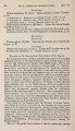
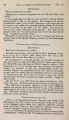
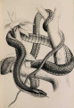
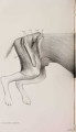
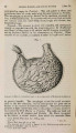
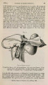
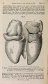
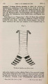
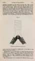
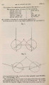
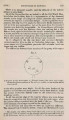
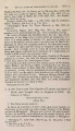
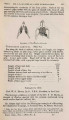
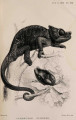
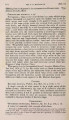
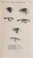
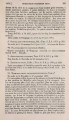
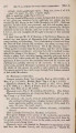
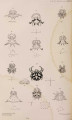
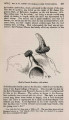
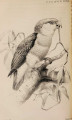
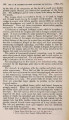
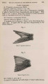
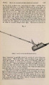
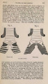
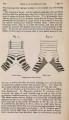
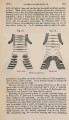
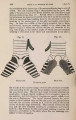
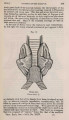
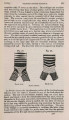
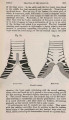
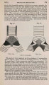
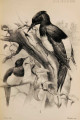
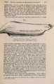
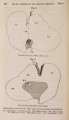
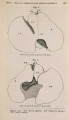
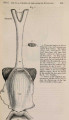
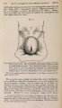
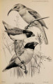
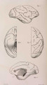
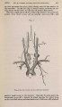
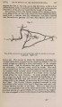
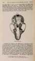
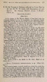
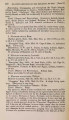
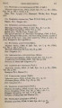
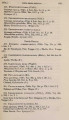
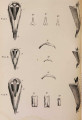
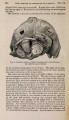
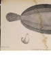
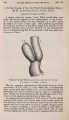
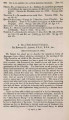
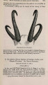
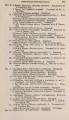
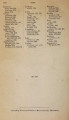
Show 1879.] MR. W. OTTLEY ON THE GROUND-HORNBILL. 465 it is continued through this plexus and divides almost immediately into two branches (14, 15), both of which supply the contents of the orbit, and eventually anastomose with the ethmoidal artery; 14 runs near the roof of the orbit, 15 under the optic nerve. The orbital plexus (17) furnishes two small branches (12, 13) to the eyelids and the muscles of the eye, a vessel (16) which runs in front of the quadrate bone, and ends in the muscles attached to the mandible, and a descending branch (8'), the course of which will be presently described. Fig. 2. Plan of the arteries for the supply of the head and neck. Right side. The vessel 21 is the internal carotid ; opposite the mark x a large offset is sent to the maxillary plexus (M.P1.), which is joined on its way by a communication from the internal maxillary artery (7). After giving off this large branch the internal carotid continues its tortuous course through a special bony canal till it reaches the interior of the cranium. A small nerve (a branch of the facial) crosses the internal carotid artery on its outer side where the communicating offset leaves that vessel. . The next branch of the vertebral is the internal maxillary (7), a large vessel which runs above the internal pterygoid muscle. Its first branch (9) ends in muscular offsets. The next (8) emerges from behind the triangular tendon of the external pterygoid, is joined by a communication from the orbital PROC. ZOOL. Soc-1879, No. XXX. 30 |