| OCR Text |
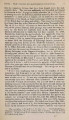
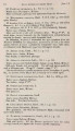
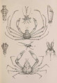
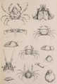
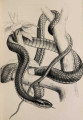
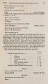
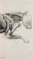
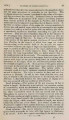
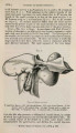
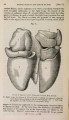
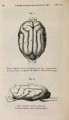
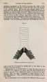
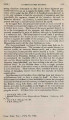
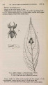
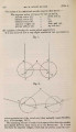
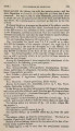
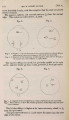
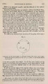
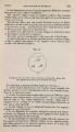
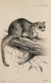
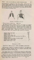
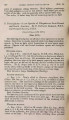
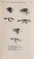
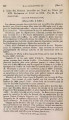
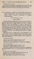
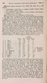
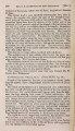
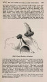
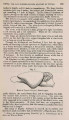
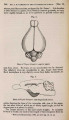
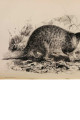
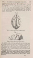
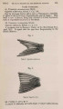
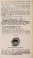
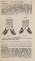
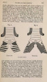
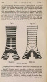
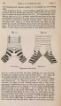
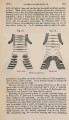
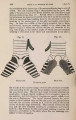
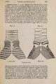
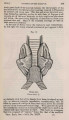
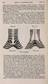
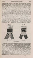
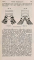
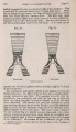
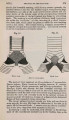
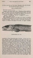
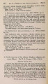
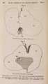
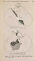
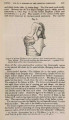
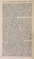
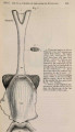
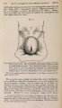
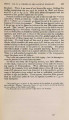
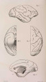
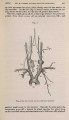
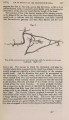
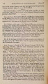
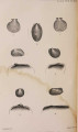
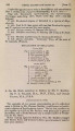
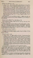
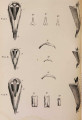
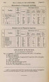
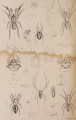
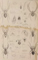
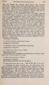
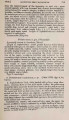
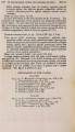
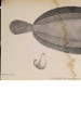
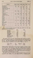
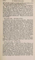
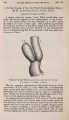
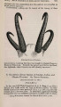
Show 1879.] EYE-MUSCLES OF MAMMALS. 123 bined, not with the inferior, but with the superior rectus ; and same disposition is found in all the Reptiles and Birds that I have examined. Not only so, but in some Mammalia, particularly those in which the eyes are placed at the side of the head, as in the Rodents and others, the muscles must be combined as they are in the fish or bird. Professor Struthers, in a paper on the action of the oblique muscles (Monthly J. of Med. Science, Oct. 1849), has already drawn attention to the differences in the direction of these muscles which are found in the Mammalia, and has pointed out that the more the eyes are directed outwards, the more does the angle which the superior oblique makes with the visual axis tend to become acute. The accompanying diagrams will explain this change in the angle. Fig. 1 represents the visual axes V A V A' parallel as in man ; S O SO'the direction of the superior oblique; the angle S c A is obtuse. In fig. 2 the axes are divergent, as in the Rabbit : the letters correspond ; the angle S c A is acute. It will be noticed also that S O SO' are directed to the front of the eye instead of to the back. This forward position of the superior oblique muscle, however, as will be presently shown, is not peculiar to those animals in which the eyes diverge. Among the Quadrumana I have examined the attachment of the eye-muscles in the following genera and species :- Fam. Simiidae.-Simla satyrus. Fam. Cercopithecidae.-Semnopithecus leucoprymnus, Cercopithecus callitrichus, C. dlbigularis, Cercocebus fuliginosus, Macacus inuus, Cynocephalus porcarius. Fam. Cebidae.-Ateles ater and A. melanochir, Mycetes seniculus, Cebus capucinus, C. hypoleucus, Nyctipithecus felinus, Saimaiis sciurea. Fam. Hapalidae.-Hapale penicillata, Midas rosalia. And in the Lemures, fam. Lemuridae, Lemur, sp.?; fam. Nycti-cebidae, Nycticebus tardigradus. In the human eye m y observations agree with Sappey's description rather than with that of Henle; and I therefore give the measurements to be found in Sappey's ' Anatomie Descriptive,' and a diagram, to serve as a standard of reference. The superior rectus is inserted -fa inch from corneal edge. It is curved ; and its outer is further from the cornea than is its inner edge. The inferior rectus at a distance of -fa. (It is also oblique like the superior.) The external rectus fa. The internal or median rectus fa to -fa. The superior oblique £# (I should rather say -fa) from the optic nerve. The inferior oblique fa from the nerve-entrance. The line of its insertion, if prolonged, would meet the optic nerve. Neither of these authors refers to the curvature of the line of insertion of the superior oblique. In Simla satyrus it will be seen that the attachments resemble |