| OCR Text |
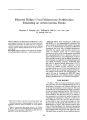
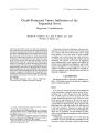
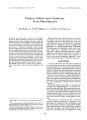
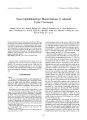
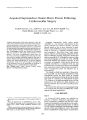
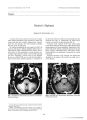
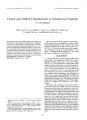
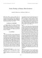
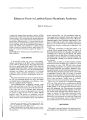
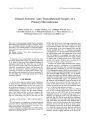
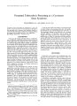
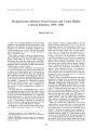
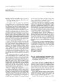
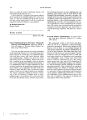
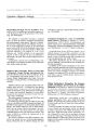
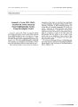
Show Journal of Nairo- Ophlhalmology 17( 3): 170- 177, 1997. 1997 Lippincott- Raven Publishers, Philadelphia Occult Perineural Tumor Infiltration of the Trigeminal Nerve Diagnostic Considerations Martin W. ten Hove, M. D., Joel S. Glaser, M. D., and Norman J. Schatz, M. D. Cutaneous carcinomas of the face, and some nasopharyngeal carcinomas, may present with facial dysesthesias and/ or facial nerve palsies in the absence of visible masses. Even with frank ophthalmoplegia, occult tumors that present in this matter may elude detection, for which reason specific diagnostic studies must be employed. We report seven cases of trigeminal nerve infiltration by occult tumors ( five squamous cell carcinomas, one basal cell carinoma, and one adenoid cystic carcinoma), and outline the clinical course, diagnostic investigations, and the subsequent management of these patients. The importance of establishing an early diagnosis before the tumor has transgressed the basal foramina is emphasized, as tumor infiltration of the cavernous sinus carries a more guarded prognosis. The use of magnetic resonance imaging to identify involved peripheral nerve branches that may then be biopsied is suggested. The pathophysiological mechanisms of neurotropic spread of tumor are reviewed. Key Words: Trigeminal nerve- Tumor- Infiltration- Cavernous sinus- Magnetic resonance imaging. Cutaneous carcinomas of the face, and some nasopharyngeal carcinomas, without obvious mass lesions may present with facial dysesthesias and/ or facial nerve palsies. Even at the stage of frank ophthalmoplegia, such occult tumors may elude detection, for which reason specific diagnostic studies must be employed. We present seven cases of trigeminal nerve infiltration by occult tumors, and outline the clinical course, diagnostic investigations, and the subsequent management of these patients. The use of magnetic resonance imaging ( MRI) to identify involved peripheral nerve branches that may then be biopsied is suggested. CASE REPORTS The clinical details for patients are outlined in Tables 1- 3. Exemplary cases are described to emphasize typical temporal profiles. Manuscript received January 11, 1996. From the Department of Ophthalmology ( M. W. t. H.), Queen's University Kingston, Ontario, Canada; and the Mercy Neurological Institute ( J. S. G., N. J. S.), Mercy Hospital, University of Miami, Miami, Florida, U. S. A. Address correspondence and reprint requests to Dr. Martin W. ten Hove, Department of Ophthalmology, Queen's University, 76 Stuart Street, Kingston, Ontario K7L 3N6, Canada. This material was presented at the Bascom Palmer Eye Institute Alumni Day, Miami, June 1994. Case 1 A 47- year- old woman presented with painful left facial paresthesias, diplopia, and left ptosis, all progressing over at least the previous year. Examination revealed acuities of 20/ 20 in each eye ( OU) with normal visual fields and color vision. There was 4 mm of left ptosis, and ductions of the left eye were severely diminished in all directions, with no mechanical restriction as determined by force ductions. The right pupil was 3 mm and reactive; the left pupil was 6 mm and unreactive. Sensation of pain and light touch was absent in the left VI and V2 distributions. There was no proptosis, and the findings on globe, orbit, and neurological examinations were otherwise unremarkable. Computerized tomography ( CT) of the head did not detect any mass, but did show asymmetry of the cavernous sinuses and bony erosion of the dorsum 170 OCCULT PERINEURAL TUMOR INFILTRATION 171 TABLE 1. Patient presentation Case 1 2 3 4 5 6 7 Age/ Sex 47, F 82, M 53, F 27, F 50, M 76, M 69, F Tumor Adenoid cystic Squamous cell Squamous cell Squamous cell Squamous cell Basal cell Squamous cell Primary site Unknown Left cheek Right lower lid Nasopharynx Left forehead Left neck and car Tip of nose Paresthesias Crawling Crawling, numbness Throbbing Numbness, pain Sand in the eye Numbness Numbness sellae. MRI confirmed fullness of the left cavernous sinus and defined a mass extending through the foramina ovale and rotundum ( Fig. 1). The findings of endocrine and hematological studies, carotid angiography, and a " blind" nasopharyngeal biopsy were normal. Sarcoid infiltration was diagnosed provisionally on the basis of gallium uptake in the hilar nodes and a single giant cell seen in bronchial washings. A course of prednisone was initiated without benefit. The facial paresthesias were improved with carba-mazepine administration. Biopsy of the cavernous sinus was recommended, but refused by the patient. Over the next 2 years, the patient developed aberrant regeneration of the left third nerve and mandibular division ( V3) weakness. Four years after onset, progressive loss of vision in the left eye was documented at 20/ f00, whereas the right remained at 20/ 20. There was a left superior altitudinal field loss. A repeat CT showed a mass extending into the posterior orbit through the superior orbital fissure. Orbital biopsy revealed adenoid cystic carcinoma in the form of malignant epithelioid cells invading the perineurium of the orbital nerves ( Fig. 2). In combination with gammaknife radiosurgery, conventional external beam radiation was delivered to cover the entire globe, orbit, cavernous sinus, and base of skull. At 6- month follow- up, the cranial nerve palsies were unchanged, but vision had decreased to 20/ 300. Case 2 An 82- year- old man had a f- year history of crawling paresthesias and numbness of the left cheek. Over previous 6 months, the numbness spread to his entire left face, and diplopia, tearing, and left ptosis had recently developed. He described difficulty chewing, with a 6- lb ( 2.7- kg) weight loss. Six months previously, a presumed squamous cell carcinoma on the left cheek had been treated by cryotherapy. Visual acuities were 20/ 100 right eye ( OD) ( prior retinal detachment) and 20/ 100 left eye ( OS). Pupils were equal and reactive, with no afferent defect. Motility was normal for the right eye, but severely limited in all directions in the left eye. Forced ductions of both globes were normal. The left cornea had a total epithelial defect secondary to neurotrophic keratopathy. Sensation was absent in all divisions to the left trigeminal nerve, including the corneal reflex. There was weakness of the left lower face. All other cranial nerve functions were normal. T2- weighted MRI showed a mass filling the cavernous sinus and the Meckel cave. The mass enhanced with gadolinium ( Gd)- DPTA, as did the left infraorbital nerve coursing through the infraorbital groove ( Fig. 3). Left pterygoid and temporalis muscles were atrophied. Biopsy of the infraorbital nerve was recommended, but the patient's debilitated state influenced the decision to irradiate the cavernous sinus and infraorbital nerve for palliation without histological confirmation. TABLE 2. Cranial nerve involvement and biopsy sites Case Cranial nerves involved Potential biopsy site L: VI, V2, V3, III VII, pupil L: VI, V2, V3, III VII R: VI, V2, VII IV, VI, Cavernous sinus", obit'w VI, L: VI, V2, V3, III, IV, VI, VII L: VI, V2, III, VII L: VI, V2, VI L: VI, V2, V3, VI Infraorbital nerve", cavernous sinus" Infraorbital nerve"'', cavernous sinus" Cavernous sinus", nasopharynx',, c Supraorbital nerve6 Cavernous sinus" Infraorbital nerve"'* " Diagnosis actually confirmed by biopsy of this site. b Biopsy site identified by magnetic resonance imaging with gad-olinium- DPTA. ' Biopsy site identified by computerized tomography. Case TABLE 3. Treatment and course Treatment Follow- up 1 XRT and gammaknife 2 XRT 3 XRT 4 XRT and chemotherapy 5 XRT 6 XRT 7 XRT 3 Months, no recurrence 1 Months, no recurrence Recurrence @ 6 months in VI 12 Months, no recurrence 14 Months, no recurrence No follow- up 18 Months, no recurrence XRT, x- ray radiation treatment. J Neuro- Ophthalmol, Vol. 17, No. 3, 1997 172 M. W. TEN HOVE ET AL. FIG. 1. Case 1. A: T1- weighted MRI with Gd- DPTA, coronal sections. B: Arrows indicate tumor in left cavernous sinus and foramen ovale. C: T1- weighted MRI, with Gd- DPTA, axial section. Case 3 A 53- year- old woman complained of right severe throbbing facial paresthesias for the previous 6 months. Three precancerous lesions had been removed from the right lower lid and right bridge of nose. She had also noted a decreased blink and facial flattening of the right side. Carbamazepine administration was unsuccessful in alleviating the paresthesias. Visual acuity was 20/ 20. The right blink was noticeably reduced, and there was some flattening of the right nasolabial fold. There was diminished sensation to pain over the right forehead and cheek. MRI revealed a mass involving the gasserian ganglion in the Meckel cave. In this case, however, the trigeminal nerve divisions did not enhance with gadolinium, nor were they appreciably enlarged ( Fig. 4). On the basis of the clinical findings, though, exploration and biopsy of the right infraorbital nerve were performed, revealing infiltration of the nerve and perineural space with squamous cell carcinoma ( Fig. 5). Conventional radiation therapy was delivered to the entire second division of the trigeminal nerve, including the gasserian ganglion. The paresthesias subsided for 6 months, subsequently returning in the distribution of the first division of the trigeminal nerve. Recurrent squamous cell carcinoma above the right brow was again confirmed by superficial nerve biopsy and resected. There has been is no clinical progression over the past 2 years. DISCUSSION The centrifugal spread of certain cutaneous cancers along cranial nerves has been well documented ( 1- 5). J Neuro- Ophthalmol, Vol. 17, No. 3, 1997 OCCULT PERINEURAL TUMOR INFILTRATION 173 FIG. 2. Case 1. Adenocystic carcinoma with malignant cells surrounding an orbital nerve. Asterisks indicate nerve fiber fasicles adjacent nests of tumor cells ( hematoxylin- eosin, X100). The first instance of perineural infiltration by tumor was reported in 1842 by Cruveilheir ( 6), and in 1963 Ballantyne et al. ( 1) reported their series of 80 cases that still stands as the largest series in the literature. Epithelial carcinomas of skin or mucus membranes are the commonest tumors to spread via the potential perineural spaces; however, nasopharyngeal tumors, lymphomas, melanoma, neurinoma, and neurofibroma may also use this route ( 4,7,8). Neurotropic spread was first felt to occur along " perineural lymphatics," a notion long disproved ( 9). This propensity for tumors to progress through perineural or endoneural fascial planes is arguably attributed to the lower resistance to invasion of these potentially distensible spaces, as compared with more direct invasion of the the nerve fibers ( 10). Thus, perineuria provide a direct pathway from the most distal cutaneous nerve branches to the proximal subarachnoid space of the central nervous system. It has been suggested that the rich vascular environment of the perineurium may provide growth factors that further promote this mode of spread ( 10). Segmental infarction of nerve branches may result from tumor infiltration of these same vasa nervosum ( 7) or, rarely, from expanding tumor within confined spaces, leading to pressure necrosis. In a large series of epithelial carcinomas, 2.5% of 807 squamous cell carcinomas, and 1% of 1,686 basal cell carcinomas, demonstrated perineural or endoneural infiltration ( 10). Thus, although a rare mode of spread, any extirpation of tumor mandates careful frozen- section examination of adjacent peripheral nerve branches. Purely neuraxial spread of up to 14 cm without contiguous tissue involvement has been reported ( 7), as have nerve branch " skip lesions" ( 11). Data on the prevalence of perineural spread of facial carcinomas to the cavernous sinus are limited. In the Thomas and Yoss review of 102 cases of para-sellar syndrome ( 12), 19 ( 20%) were due to nasopharyngeal carcinomas infiltrating the cavernous sinus via cranial nerves, whereas none were due to cutaneous squamous or basal cell carcinoma. The symptoms of dysesthetic pain are described as throbbing, stabbing, burning, stinging, or crawling. Some patients ( cases 1- 5 inclusive) presented with facial weakness, which indicates tumor infiltration of peripheral branches of the seventh nerve. Often these early signs do not lead to appropriate investigation or diagnosis until other cranial nerves become involved, as in all of our cases. The " numb chin syndrome" refers to isolated mental branch neuropathy resulting from infiltrative lymphoma ( 13- 15), squamous cell carcinoma ( 2), or metastatic lung, breast, and renal cell carcinoma ( 14- 16). The radiological assessment of such suspected lesions in patients includes coronal and axial CT views of the basal foramina. An asymmetry of > 2 mm of the foramen rotundum or > 4 mm of the foramen ovale suggests pathological involvement, but may not adequately delineate an optimal biopsy site ( 17). J Neuro- Ophlhalmol, Vol. 17, No. 3, 1997 174 M. W. TEN HOVE ET AL. FIG. 3. Top: Case 2. T1- weighted MRI with Gd- DPTA, axial section. Thick arrows indicate a mass filling Meckel's Cave ( characteristically seen as a void, Curved arrow). The trigeminal nerve root shows enhancement as its heads toward the Gasserian ganglion ( Thin arrows). Bottom: T1- weighted MRI, with Gd- DPTA, coronal section. Enhancement of left infraorbital nerve in similar case 5. J Neuro- Ophllmlmol, Vol. 17, No. 3, 1997 OCCULT PERINEURAL TUMOR INFILTRATION 175 FIG. 4. Case 3. T1- weighted MRI, coronal section showing diffuse mass in the right cavernous sinus { arrows) with Gd- DPTA, enhancement of the mass. Thin- section axial and coronal Tl- weighted MRIs, before and after administration of Gd- DPTA, are the optimal images for detecting changes of the trigeminal nerve from the Meckel cave to peripheral nerve infiltrations ( 18). The use of a fat- suppression technique may be a useful adjunct to conventional MRI sequences ( 19). Laine's series ( 20) of seven cases with tumor infiltration of the trigeminal nerve led him to suggest the following radiological clues: ( a) an enlarged foramen ovale; ( b) smooth, isointense enlargement of the second or third division of the trigeminal nerve, ( c) bulging of the lateral wall of the cavernous sinus; ( d) replacement of the normally hypointense cistern of the gasserian ganglion; and ( e) atrophic changes in the masticatory muscles. To our knowledge, we present the first instance ( case 2) of perineural infiltration of the infraorbital FIG. 5. Case 3. Squamous cell carcinoma infiltrating the perineural space ( arrows) of the right infraorbital nerve ( hematoxylin-eosin, X100). .1 " *- •*. • * ^ .' V""; i ; > > V _ ; A;. ^- -^ » -.-* r • 4 / * i 776 M. W. TEN HOVE ET AL. nerve detected with Gd- DPTA. Although infiltration of infraorbital and supraorbital nerves can produce an enlarged infraorbital foramen or supraorbital notch ( 1,4,7), this latter sign evolves only after significant spread has occurred. Gulya et al. ( 21) have reported a case with Gd- DPTA enhancement of the gasserian ganglion and atrophy of the masticatory muscles ipsilateral to a primary cutaneous squamous cell carcinoma. While Gd- DPTA enhancement of peripheral nerves is not yet generally recognized, enhancement of the oculomotor nerve has been demonstrated in lymphoma, leukemia, viral meningitis, neurofibromatosis, HIV polyneuropathy, migraine, Tolsa- Hunt syndrome, and diabetic ophthalmoplegia ( 22). If an involved peripheral nerve can be detected by the criteria just described, an open biopsy of the involved nerve segment is indicated. Indeed, if the clinical picture suggests perineural tumor infiltration, even in the absence of radiological nerve changes, an exploration may still prove positive, as in case 3. Excision of a small portion of an already infiltrated nerve will not substantially increase the clinical neurological deficit ( 8). With tumor involvement of the cavernous sinus, a transcranial biopsy may be considered. The risk of venous hemorrhage is minimal when phlebographic imaging demonstrates occlusion of the cavernous sinus ( 8,23). Alternatively, CT- guided fine- needle aspiration techniques through the foramen ovale have been used with some success to verify perineural spread ( 24). In our series, two instances apparently arose de novo ( case 1 and 4), without peviously detected lesions of skin or nasopharynx. The remaining cases evolved in areas where primary skin lesions had occurred. Following extirpation of such lesions, delayed perineural infiltration may occur from within a few months to several years. Although the majority of these tumors are indolent, few cases are actually detected before the tumor has transgressed the base of skull. Therefore, the prognosis for symptomatic control is variable. Biopsy may itself afford relief from dysesthetic symptoms ( 8). Painful dysesthesia may also respond to external beam radiation or to radiosurgery. Subsequent aberrant regeneration of either the trigeminal or the facial nerve may occur, as in case 1. The prognosis for survival is a function of tumor type, degree of anaplasia, and extent of spread. Cases diagnosed prior to intracranial involvement, and when the entire extracranial portion of the involved nerve may be included within the field of radiation, invariably have the better prognosis. The presence of paresthesia and dysesthesia in the distribution of the trigeminal nerve should suggest the possibility of tumor infiltration. Involvement of V3 is associated with trigeminal motor dysfunction. Cavernous sinus invasion occurred in five of our seven cases, and two patients had focal areas of facial weakness implying infiltration of the facial nerve. The most common pattern of perineural spread was along the sensory peripheral nerve branches to the gasserian ganglion. Spread in both anterograde and retrograde directions was seen in one patient, with sensory symptoms progressing from V2 to Vi ( case 3). Biopsy of accessible peripheral nerve branches supplying symptomatic areas can be a practical means of establishing a tissue diagnosis. Such biopsies are associated with significantly less morbidity than either open biopsies of the cavernous sinus or transforaminal CT-guided biopsies. MRI may be extremely helpful in identifying peripheral biopsy sites, but the decision for exploratory biopsy may be made strictly on clinical grounds. Acknowledgment: M. W. t. H. received financial support from the McLaughlin Foundation, Toronto, Canada. REFERENCES 1. Ballantye AJ, McCarlcn AB, Ibanez ML. The extension of cancer of the head and neck through peripheral nerves. Am J Surg 1963; 106: 651- 67. 2. Brazis PW, Volger JB, Shaw KE. The " numb cheek- limp lower lid" syndrome. Neurology 1991; 41: 327- 8. 3. Clousten PD, Sharpe DM, Corbett AJ, Kos S, Kennedy PJ. Perineural spread of cutaneous head and neck cancer. Arch Neurol 1990; 47: 73- 7. 4. Moore CE, Hoyt WF, North JB. Painful ophthalmoplegia following treated squamous cell carcinoma of the forehead. Med J Aust 1976; 1: 657- 9. 5. Trobe JD, Hood CI, Parsons JT, Quisling RG. Intracranial spread of squamous carcinoma along the trigeminal nerve. Arch Ophthalmol 1982; 100: 608- 11. 6. Cruveilhier J. Maladies des nerfs: anatomic pathologique du corps humain. Paris: JB Baillicro, 1835- 1842 ( Pari 2, 1935): 3. 7. Dodd GD, Dolan PA, Ballantye AJ, Ibanez ML, Chau P. The dissemination of tumors of the head and neck via the cranial nerves. Radiol Clin North Am 1994; 8: 445- 61. 8. Unsold R, Safran AB, Safran E, Hoyt WF. Metastatic infiltration of nerves in the cavernous sinus. Arch Neurol 1980; 37: 59- 61. 9. Larson DL, Rodin AE, Roberts DK. Perineural lymphatics: myth or fact? Am J Surg 1966;! 12: 488- 92. 10. Mohs FE, Lathrop TG. Modes of spread of cancer of the skin. Arch Dermatol 1952; 66: 427- 39. 11. Cottel WI. Perineural invasion by squamous- cell carcinoma. J Dermatol Surg Oncol 1982; 8: 589- 600. 12. Thomas JE, Yoss RE. The parascllar syndrome: problems in determining etiology. Mayo Clin Proc 1970; 45: 617- 23. 13. Horton J, Means ED, Cunningham TJ, Olson KB. The numb chin in breast cancer. / Neurol Neurosurg Psychiatry 1973; 36: 211- 6. 14. Kuntzer T, Borgousslavsky J, Rilliet B, Uldry PA, Tribolet N, Regli F. Herald facial numbness. Eur Neurol 1992; 32: 297- 301. 15. Massey EW, Moore J, Schold SC. Mental neuropathy from systemic cancer. Neurology 1981; 31: 277- 81. 16. Williams HM, Diamond HD, Craver LF, Parsons H. Neurological complications of lymphomas and leukemias. Springfield, IL: Charles C Thomas, 1959: 4- 8. 17. Medina JE, Pavlovich A, Wilson DA. Magnetic resonance imaging in the diagnosis of intracranial spread of carcinoma via the trigeminal nerve. Otolaryngol Head Neck Surg 1990; 102: 417- 9. 18. Parker GD, Harnsberger HR. Clinical- radiologic evaluation of trigeminal neuropathy. Semin Ultrasound CT MR 1987; 8: 214- 39. J Neuro- Oplulmlmol, Vol. 17, No. 3, 1997 OCCULT PERINEURAL TUMOR INFILTRATION 177 19. Majoie CB, Hulsmans FH, Castelijns JA, Walter A, Bras J, Peetcrs FL. Perineural tumor extension of facial malignant melanoma: CT and MRI. J Compul Assist Tomogr 1993; 17: 973- 5. 20. Laine FJ, Sen ME, Braun IF, Nadel L, Som PM. Perineural tumor extension through the foramen ovale: evaluation with MR imaging. Radiology 1990; 174: 65- 71. 21. Gulya AJ, Scher R, Schwartz AJ, Wilson WR. Facial and trigeminal neural dysfunction by a primary cutaneous squamous cell carcinaoma: MRI and clinicopathological correlates. Am J Otol 1994; 13: 587- 90. 22. Mark AS, Blake P, Atlas SW, Ross M, Kolsky M. Gd- DTPA enhancement of the cisternal portion of the oculomotor nerve on MR imaging. AJNR 1992; 13: 1463- 70. 23. Sekhar LN, M0ller AR. Operative management of tumors involving the cavernous sinus. J Neurosurg 986; 64: 879- 89. 24. Barkos JA, Dilton WP. Lesions of the foramen ovale: CT guided fine needle aspiration. Radiology 1992; 182: 573- 5. J Neuro- Ophthalmol, Vol. 17, No. .?, 1997 |