| Title |
Temporal Crescent Syndrome with Magnetic Resonance Correlation |
| Creator |
Chavis, PS; al-Hazmi, A; Clunie, D; Hoyt, WF |
| Affiliation |
Department of Ophthalmology, King Khaled Eye Specialist Hospital, Riyadh, Saudi Arabia. |
| Abstract |
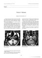
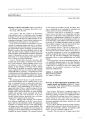
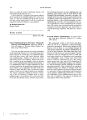
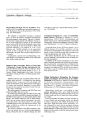
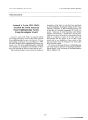
BACKGROUND: A young woman with a history of controlled hypertension noted a suddenly decreased peripheral temporal field in the left eye. This occurred after moderate peripartum hypertension. METHOD: A monocular peripheral temporal crescentic defect could be plotted on Goldmann visual fields despite a normal dilated peripheral retinal examination and normal disc appearance. RESULT: A dilated parieto-occipital sulcus could be seen on computed tomography, and magnetic resonance imaging showed changes consistent with atrophy and gliosis in the cuneus, precuneus, and anterior calcarine cortex surrounding the parieto-occipital sulcus. CONCLUSION: By magnetic resonance imaging, this can be seen to comprise less than 10% of the visual cortex, as suggested by the Horton and Hoyt revised Holmes map. The temporal crescent syndrome is a rare monocular retrochiasmatic visual field defect that can be correlated to a lesion along the parieto-occipital sulcus. |
| Subject |
Adult; Blood Pressure; Brain/pathology; Brain Diseases/complications; Brain Diseases/diagnosis; Female; Hemianopsia/etiology; Humans; Hypertension/complications; Magnetic Resonance Imaging; Syndrome; Visual Acuity; Visual Fields |
| Format |
application/pdf |
| Publication Type |
Journal Article |
| Collection |
Neuro-Ophthalmology Virtual Education Library: Journal of Neuro-Ophthalmology Archives: https://novel.utah.edu/jno/ |
| Publisher |
Lippincott, Williams & Wilkins |
| Holding Institution |
Spencer S. Eccles Health Sciences Library, University of Utah |
| Rights Management |
© North American Neuro-Ophthalmology Society |
| Setname |
ehsl_novel_jno |
| ID |
224824 |
| Reference URL |
https://collections.lib.utah.edu/ark:/87278/s6962pmg/224824 |