| OCR Text |
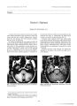
Show Journal of Neuro- Ophthalmology 17( 3): 162- 165, 1997. © 1997 Lippincott- Raven Publishers, Philadelphia Anterior Ischemic Optic Neuropathy and Activated Protein C Resistance A Case Report and Review of the Literature Bradford B. Worrall, M. D.,* Golnaz Moazami, M. D.,| Jeffery G. Odel, M. D. j and Myles M. Behrens, M. D. f Nonarteritic anterior ischemic optic neuropathy ( AION) is a well- described entity that is believed to be caused by abnormal anatomy of the optic disc and to be precipitated by several stressors or disease states. Activated protein C resistance ( APCR) is a recently described mutation of factor V ( FV) gene that renders FV resistant to cleavage by activated protein C. APCR predisposes to thrombotic events. The case of a 61- year-old woman with AION and activated protein C resistance is presented, and the management is discussed. We thoroughly review the literature on these two conditions. We propose that a prospective analysis of the potential role of APCR in some cases of AION is needed and suggest that physicians consider this and other prothombotic states when evaluating patients with AION. Key words: Anterior ischemic optic neuropathy- Activated protein C resistance- Thrombophilia. Anterior ischemic optic neuropathy ( AION) classically occurs as sudden painless loss of vision in one eye rendering an altitudinal ( arcuate) or less commonly cen-trocecal field defect ( 1). In 40% of patients, subsequent loss of vision in the other eye ensues ( 2), but recurrent attacks in the same eye are far less common ( 3). The underlying mechanism is thought to be ischemic injury to a crowded ( small cup) optic nerve ( 4). AION has been reported with a higher than expected frequency in patients with diabetes, hypertension, and ischemic heart disease ( proatherosclerotic states; ( 5,6,7)); in cases of significant blood loss ( 8); and in other systemic disorders predisposing to ischemia ( 9). Acheson and Sanders describe a series of patients with AION who have other potentially procoagulant conditions ( 10), but no one to our knowledge has described a case of AION associated Manuscript received September 18, 1996; accepted January 13, 1997. From the * Departments of Neurology and tOphthalmology/ Division of Neuro- Ophthalmology, Columbia Presbyterian Medical Center, New York, New York. Address correspondence and reprint requests to Dr. Bradford B. Worrall, Neurological Institute, Box 28, Columbia Presbyterian Medical Center, 710 West 168th Street, New York, NY 10032- 2603. with activated protein C resistance ( APCR), one of the most recently reported prothrombotic abnormalities ( 11). We report such a case here. CASE REPORT A 61- year- old woman noticed abrupt and painless loss of vision in her right eye ( OD) on awakening that worsened the following day. She had no jaw claudication, temporal tenderness, polymyalgia rheumatica, history of transient visual loss, or other neurologic symptoms. She had no history of diabetes, hypertension, migraine, or abnormal clotting. She was on a regimen of one aspirin tablet ( 325 milligrams) a day. Family history is negative for similar visual problems or for thrombophilia. On examination on the fifth day, her visual acuity was 20/ 20 in each eye ( OU) with 6/ 6 on American Optical/ Hardy Rand color plates OU. She had a right relative afferent pupillary defect reversed by 0.6 log unit neutral density filter. Fundoscopic examination revealed a pale, swollen disc with splinter hemorrhages OD. There was some pallor without swelling and absence of physiologic cup on the left ( OS). Visual fields revealed an inferior altitudinal defect OD and a suggestion of a mild paracentral defect OS. Results of the neurologic examination were otherwise normal. Erythrocyte sedimentation rate was elevated ( 60 mm/ hour). Results of the temporal artery biopsy were negative for giant cell arteritis. Magnetic resonance imaging of head and orbits gave negative findings. She was diagnosed with acute nonarteritic AION OD and a prior AION OS. Extensive evaluation for any underlying process found a decreased ratio of activated partial thromboplastin time ( APTT) in presence of activated protein C ( APC) over the APTT consistent with APCR. We proposed that her hypercoagulable tendency may have had a role in her AION and considered long-term anticoagulation therapy. An exhaustive review of the literature did not reveal known cases or clear guidelines for therapy. Eventually, based on current recommendations for other procoagulant conditions, no anticoagulation was initiated, and she remained on aspirin. 162 AION AND ACTIVATED PROTEIN C RESISTANCE 163 LITERATURE REVIEW AND DISCUSSION During the last 3 years, several new discoveries in molecular mechanisms of the coagulation-anticoagulation system have enhanced our understanding of thrombophilia. Dahlback and others in 1993 found resistance to APC in two families with hereditary hypercoagulability ( 12). They subsequently identified this abnormality in approximately 40% of consecutive patients referred for unexplained thrombosis, although the incidence in the general population is 7%. APCR was therefore recognized as a major cause of primary thrombophilia ( 13). Bertina and others then identified a point mutation in the factor V ( FV) gene that results in a specific amino acid substitution in FV ( 14) that was shown to segregate with APCR ( 15). Currently, this mutation is thought to alter the binding site for APC to cleave and inactivate FV. The coagulation cascade and the natural anticoagulants maintain a balance during steady state to prevent spontaneous bleeding and blood loss and to prevent spontaneous clotting and interruption of blood flow. In the setting of vascular injury, the coagulation cascade stops blood loss and the natural anticoagulants limit the clot formation to sites of injury. Protein C is a vitamin K- dependent protein synthesized by the liver and is part of a potent anticoagulation system. Thrombin in the serum binds to thrombomodulin, a receptor on the endothelial membranes. This complex changes conformation to allow binding and cleavage of protein C to form APC. Protein S is a crucial cofactor for APC. In normal serum, the APC- protein S complex binds to activated FV and activated factor VIII ( FVIII) and degrades them to inactive forms. This arrests the coagulation cascade and inhibits the formation of fibrin ( 16,17). People with APCR are thought to have a mutation that changes the binding site on FV and prevents the APC-protein S complex from attaching and degrading FV to an inactive form. This mutation appears to have no effect on the procoagulant properties of FV ( 18). Thus, the normal down- regulation of the coagulation cascade is less efficient in people with APCR. Heterozygotes retain some APC activity whereas homozygotes have no FV with APC binding sites. Both heterozygotes and homozygotes are at increased risk of thrombotic events, especially when facing a disturbance to the homeostatic condition. Rosendaal and coworkers postulated that the risk for thrombosis was 80 times greater for homozygotes than the accepted risk for the general population, giving them an annual absolute risk of 1 % to 2% per year ( 19). APCR can be identified by several methods. The most precise is identifying the substitution of glycine ( Gly) for arginine at position 506 of FV or the guanine for alanine at position 1691 on the FVgene ( 14,20). However, this is impractical clinically. The APC sensitivity ratio ( APC-SR) can be measured using commercially available kits. The APC- SR is a ratio of the APTT in the presence APC to the APTT alone. It has been shown to be an accurate and reproducible way to identify APCR ( 21,22). The test is logistical ly simple, requiring aliquots of APC to be mixed with the patient's serum in the presence of calcium chloride. The prolongation of the APTT results from the APC's anticoagulant properties. In patients with APCR, no prolongation of the APTT occurs and the ratio remains low ( 23). Various reports claim association of APCR with certain pathologic conditions: spontaneous deep venous thrombosis ( DVT) ( 19,24), DVT with oral contraceptives ( OCP; 25), pregnancy complications ( 26), spontaneous and perioperative arterial thrombosis ( 27,28), myocardial infarction ( 29,30), and cerebral infarction ( 31- 34). Not all authors find such associations, especially with regard to coronary artery disease ( 35,36). Several authors point out the low incidence of thrombotic or thromboembolic complications in patients who are heterozygous for the FV mutation- a relatively common occurrence in the general population. Press and Goodnight found a baseline incidence of 7.6% of het-erozygocity for FV 506 Gly mutation in consecutive Red Cross Blood donors ( 23). They propose a multifactorial role where the presence of one gene for FV 506 predisposes people to hypercoagulable complications in the presence of endogenous or exogenous factors ( 23). This " two hit" hypothesis for procoagulant phenomena has been supported anecdotally and in several small studies by the coincidence of two prothrombotic states: either endogenous ( protein C deficiency, protein S deficiency, or a second gene for mutant FV; 30,37), or exogenous ( immobility, surgical intervention, OCP use; 25- 27,38,39) in patients with thrombophilia. Larsson and colleagues also discuss this phenomenon and note that although the incidence of APCR is estimated at 2% to 7% in the population, the incidence of spontaneous thrombosis is much lower. Unfortunately, their series was too small to detect the role of specific secondary factors such as cigarette abuse or OCP use ( 40). The pathophysiologic mechanism in nonarteritic AION is thought to be secondary to anatomic variations that predispose to vascular insufficiency to the optic nerve. Most of the optic nerve behind the prelaminar optic nerve head receives its vascular supply from the posterior ciliary arteries. A small cup- to- disc ratio reflects a small opening in the sclera that may predispose these supplying vessels to compromise, especially in the face of nerve fiber swelling ( 41). The symmetry of disc crowding predisposes the second eye to subsequent similar involvement in 40% or more of the cases, but the decreased crowding after an episode of AION is presumed to protect against recurrence in the same eye except in rare occasions ( 3,42). McLeod and coworkers hypothesized a relation between moderate ischemic injury to neurons and axonal transport dysfunction ( 43). This breakdown of transport leads to axonal swelling, which may, in turn, perpetuate the ischemic injury by further compromise of vascular supply. Experimentally induced ischemic injury to the optic nerve by occlusion of the posterior ciliary arteries in nonhuman primates often leads to a condition similar ./ Neuro- Ophllmlmol, Vol. 17, No. . i, 1997 164 B. B. WORRALL ET AL. to AION ( 43). A system of collaterals may protect the optic nerve in some cases. Lavin ( 42) and Beck and colleagues ( 44) suggest that a second problem, such as small vessel vasculopathy ( atherosclerotic) or age-related collateral dropout, is necessary for the full syndrome of AION. Alternatively, Hayreh contends that it is local edema and not axonal dysfunction that leads to disc swelling and neuronal death ( 45). In their series, Acheson and Sanders found that seven patients referred for evaluation of nonarteritic AION had a hypercoagulable state ( 10). Their investigation included routine screening and a hematologic evaluation including prothrombin time, APTT, lupus anticoagulant, anticardiolipin antibody, protein C, protein S, and anti-thrombin III ( AT III). They identified two patients with protein C deficiency, one with protein S deficiency, one with AT III deficiency, and three with lupus anticoagulant ( one of the three only transiently). This series raised the possibility of a role for hypercoagulability in the pathogenesis of some cases of AION ( 10). Further support for a potential role for hypercoagulability in AION comes from the reports of hemodynamic compromise of the optic nerve ( 46,47) and of amaurosis fugax ( 48) in patients with well- characterized hypercoagulable states such as antiphospholipid antibodies, anticardiolipin antibodies, and protein C deficiency. Additionally, Kleiner and others ( 49) describe lupus anticoagulant retinopathy: an occlusive vasculopathy that is a different entity but potentially has a similar mechanism. Dhote and associates identified a patient with central retinal vein occlusion ( CRVO) and APCR ( 50). Williamson and others found an association between increased blood viscosity ( the proposed mechanism of CRVO) and APCR in their series of 87 patients. The prevalence of APCR is 12% in CRVO compared with 5% in their controls ( 51). Larsson and others showed in a case-control study that 8 of 31 consecutive patients younger than 50 years of age with CRVO had APCR. They therefore deem APCR the most common known cause of CRVO ( 40). Again, this is a different disease but is especially provocative in our discussion of processes affecting the eyes. AION has been well described as associated with conditions that stress the coagulation cascade or counteract the thrombolytic system. AION occurs after significant blood loss, especially from the gastrointestinal tract ( 7), and after major surgery, especially coronary artery bypass ( 8). Both of these situations may promote proco-agulant tendencies. Given the relatively common occurrence of the FV Gly mutation in the general population, the stress of surgery or hemorrhage could be a " second hit" in a vulnerable population. The possible association between APCR and AION seems reasonable and may have implications for treatment. The current recommendation for patients with APCR ( and other potentially thrombophilic states) is to consider anticoagulation after a single thrombotic event if there are ongoing exogenous risk factors or unique circumstances, such as concurrence of two profhombotic conditions ( 16,17). Patients with a single event require treatment in such circumstances. If two thrombotic events occur, lifelong anticoagulation is indicated unless specifically contraindicated ( 16,17). Hunt recommends a short course of anticoagulation after a thrombotic event in persons identified to have a genetic predisposition to thrombophilia and then prophylactic anticoagulation for hemostatic stress such as surgery ( 52). In both the proposed procoagulant risk of APCR and the hypothetical mechanism in AION of ischemic injury, the coexistence of two factors is crucial. In considering our patient, her anatomy leaves her at risk for AION and her genetic predisposition for thrombophilia may provide sufficient loss of flow from in situ thrombosis or emboli to initiate the ischemic damage. Similarly, the FV 506 Gly gene mutation in this patient with the APCR may lead to thrombosis secondary to the intrinsic anatomical vascular compromise, abnormal flow, and stasis from her small cup- to- disc ratio ( 18). CONCLUSION Given that vision is such an integral function, can we wait for the " second event" to initiate therapy? A large study of thrombophilia and AION is necessary to determine whether the occurrence of these two phenomena is merely coincidental or implies a causal relation. If the latter is borne out, patients with AION should be evaluated for thrombophilic states. Those with a potentially prothrombotic condition may fall into the category of patients who should receive anticoagulation after a single event. Until such a study is done, physicians should consider an evaluation of procoagulant state including APCR in patients who have AION. Acknowledgment: We thank Karen Sullivan for her help in revising this manuscript. REFERENCES 1. Arnold AC, Hepler RS. Natural history of Non- arteritic anterior ischemic optic neuropathy. / Neuroophthalmol 1994; 2: 66- 9. 2. Beri M, Klugman MR, Kohler JA, Hayreh SS. Anterior ischemic optic neuropathy VII: incidence of bilaterality and various influencing factors. Ophthalmology 1987; 94: 1020- 128. 3. Borchert M, Lessell S. Progressive and recurrent non- arteritic anterior ischemic optic neuropathy. Am J Ophthalmol 1988; 106: 443- 9. 4. Feit RH, Tomsak RL, Ellenberger C. Structural factors in pathogenesis of ischemic optic neuropathy. Am J Ophthalmol 1989; 98: 105- 8. 5. Tice DA. Ischemic optic neuropathy and cardiac disease. Ann Thome Surg 1987; 44: 677. 6. Sweeny PI, Breuer AC, Selhorst IB, et al. Ischemic optic neuritis: a complication of cardiopulmonary bypass surgery. Neurology 1982; 32: 560- 2. 7. Ellenberger C Jr. Ischemic optic neuropathy as possible early complication of vascular hypertension. Am J Ophthalmol 1979; 88: 1045- 51. 8. Hayreh SS. Anterior ischemic neuropathy: clinical features and pathogenesis of post- hemorrhagic amaurosis. Ophthalmology 1987; 94: 1488- 502. 9. Hayreh SS, Joos KM, Podhajsky PA, Long CR. Systemic diseases associated with non- arteritic AION. Am J Ophthalmol 1994;] 18: 766- 80. J Neuw- Ophthulmol, Vol. 17, No. 3, 1997 AION AND ACTIVATED PROTEIN C RESISTANCE 165 10. Acheson JF, Sanders MD. Coagulation abnormalities in ischemic optic neuropathy. Eye 1994; 8( pt. l): 89- 92. 11. Bokarewa MI, Bremme K, Falk G, Sten- Linder M, Egberg N, Blombeck M. Studies on phospholipid antibodies, APC- resistance and associated mutation in the coagulation factor V gene. Thromb Res 1995; 78: 193- 200. 12. Dahlback B, Carlsson M, Svensson PJ. Familial thrombophilia due to a previously unrecognized mechanism characterized by poor anticoagulant response to activated protein C. Proc Natl Acad Sci USA 1993; 90: 1004- 8. 13. Svensson PJ, Dahlback B. Resistance to activated protein C as a basis for venous thrombosis. N Engl J Med 1984; 330: 517- 22. 14. Bertina RM, Koeleman BPC, Koster T, et al. Mutation in blood coagulation factor V associated with resistance to activated protein C. Nature 1994; 369: 64- 7. 15. Zoller B, Dahlback B. Linkage between inherited resistance to activated protein C and factor V gene mutation in venous thrombosis. Lancet 1994; 343( 8912): 1536- 8. 16. Bauer KA. Hypercoagulability: a new cofactor In the protein C anticoagulant pathway. N Engl J Med 1994; 330: 566- 7. 17. Schafer AI. Hypercoagulable states: molecular genetics to clinical practice. Lancet 1994; 344( 8939- 8940): 1739^ 12. 18. Dahlback B. Molecular genetics of thrombophilia: factor V gene mutation causing resistance to activated protein C as a basis of the hypercoagulable state. J Lab Clin Med 1995; 125: 566- 71. 19. Rosendaal FR, Koster T, Vendenbroucke JP, Reitman PH. High risk of thrombosis in patients homozygous for factor V Leiden. Blood 1995; 85: 1504- 8. 20. Dahlback B. Resistance to activated protein C, the Arg506 to Gin mutation in factor V gene, and venous thrombosis. Thromb Hae-most 1995; 73: 739^ 12. 21. Rosen S, Johansson K, Lindberg B, Dahlback B. Multicenter evaluation of a kit for activated protein C resistance on various coagulation instruments using plasmas from healthy individuals. Thromb Haemost 1994; 72: 255- 60. 22. de Ronde H, Bertina RM. Laboratory diagnosis of APC- resistance: a critical evaluation of the test and the development of diagnostic criteria. Thromb Haemost 1994; 72: 880- 6. 23. Press RD, Goodnight SH. Predisposition to thrombosis by a factor V mutation causing hereditary resistance to activated protein C. West J Med 1995; 162: 450- 2. 24. Legnani C, Palareti G, Biagi R, Coccheri S. Activated protein C resistance in deep- venous thrombosis. Lancet 1994; 343( 8896): 541- 2. 25. Vandenbroucke JP, Koster T, Briet E, Reitsman PH, Bertina RM, Rosendaal FR. Increased risk of venous thrombosis in oral-contraceptive users who are carriers of factor V Leiden mutations [ letter]. Lancet 1994; 344( 8935): 1435- 7. 26. Cook G, Walker ID, McCall F, Conkie JA, Greer IA. Familial thrombophilia and activated protein C resistance: thrombotic risk in pregnancy? Br J Hematol 1994; 87: 873- 5. 27. Lindblad B, Svensson PJ, Dahlback B. Arterial and venous thromboembolism with fatal outcome and resistance to activated protein C [ letter]. Lancet 1994; 343( 8902): 917. 28. Ma DD, Aboud MR, Williams BG, Isbister JP. Activated protein C resistance and inherited factor V mis- sense mutation in patients with venous and arterial thrombosis in a hematology clinic. Aust N ZJ Med 1995; 25: 151- 4. 29. Emmerich J, Poirier O, Evans A, et al. Myocardial infarction, Arg 506 to Gin factor V mutation, and activated protein C resistance [ letter]. Lancet 1995; 345( 8945): 321. 30. Holm J, Zoller B, Svensson PJ, Berntorp E, Erhart L, Dahlback B. Myocardial infarction associated with homozygous resistance to activated protein C [ letter]. Lancet 1994; 344( 8927): 952- 3. 31. Halmayer WM, Hausofer A, Schon R, Fischer M. The prevalence of poor anticoagulant response to activated protein C among patients suffering from stroke or venous thrombosis and among healthy subjects. Blood Coagul Fibrinolysis 1994; 5: 51- 7. 32. Forsyth PD, Dolan G. Activated protein C resistance in cases of cerebral infarction [ letter]. Lancet 1995; 345( 8952): 795. 33. Simioni P, de Ronde H, Prandoni P, Saladini M, Bertina RM, Girolami A. Ischemic stroke in young patients with activated protein C resistance: a report of three cases belonging to different kindred. Stroke 1995; 26: 885- 890. 34. Albuchner JF, Guiraud- Chaumeil B, Chollet F, Cadroy Y, Sie P. Frequency of resistance to activated protein C due to factor V mutation in young patients with ischemic stroke [ letter]. Stroke 1996; 27: 766- 7. 35. Marz W, Seydewitz H, Winklemann B, Chen M, Nauck M, Witt I. Mutation in coagulation factor V associated with resistance to activated protein C in patients with coronary artery disease [ letter]. Lancet 1995; 345( 8948): 526. 36. Samani NJ, Lodwick D, Martin D, Kimber P. Resistance to activated protein C and risk of premature myocardial infarction [ letter]. Lancet 1994; 344( 8938): 1709- 10. 37. Simioni P, Girolami A. Homozygous factor V- deficient patients show resistance to activated protein C whereas heterozygotes do not. Blood Coagul Fibrinolysis 1994; 5: 825- 7. 38. Zoller B, Bernsdotter A, Garcia de Frutos P, Dahlback B. Resistance to activated protein C as an additional genetic risk factor in hereditary deficiency of protein S. Blood 1995; 85: 3518- 23. 39. Bokarewa MI, Blomback M, Egberg N, Rosen S. A new variant of interactions between phospholipid antibodies and the protein C system. Blood Coagul Fibrinolysis 1994; 5: 37- 41. 40. Larsson J, Olafdottir E, Bauer B. Activated protein C resistance in young adults with central retinal vein occlusion. Br J Ophthalmol 1996; 80: 200- 2. 41. Beck RW, Servais GE, Hayreh SS. Anterior ischemic optic neuropathy: cup to disc ratio and its role in pathogenesis. Ophthalmology 1987; 94: 1503- 8. 42. Lavin PJM. Optic disc risk factors for non- arteritic anterior ischemic optic neuropathy. Am J Ophthalmol 1994; 117: 822. 43. McLeod D, Marshall J, Krohner EM. Role of axoplasmic transport in the pathophysiology of ischemic disc swelling. Br J Ophthalmol 1980; 64: 1488- 502. 44. Beck RW, Savino PJ, Repka MX, Schatz NJ, Sergott RC. Optic disc structure in anterior ischemic optic neuropathy. Ophthalmology 1984; 91: 1334- 7. 45. Hayreh SS. Axoplasmic transport and ischemic disc swelling. Br J Ophthalmol 1981; 65: 70- 71. 46. Watts MT, Greaves M, Clearkin LG, Malia RG, Cooper SM. An-tiphospholipid antibodies and ischemic optic neuropathy [ letter!. Lancet 355( 8793): 613- 4, 1990. 47. Ogino S, Iwamoto K, Yamamoto H, Yamaguchi K, Kondo M. A case of optic neuritis associated with anticardiolipin antibodies [ letter]. Rinsho Shikeigaku 1992; 32: 330- 2. 48. Smith DB, Ens GE. Protein C deficiency: a cause of amaurosis fugax [ letter]? J Neurol Neurosurg Psychiatry 1987; 50: 361- 2. 49. Kleiner RC, Najarian LV, Schatten S, Jabs DA, Patz A, Kaplan HJ. Vaso- occlusive retinopathy associated with antiphospholipid antibodies ( lupus anticoagulant retinopathy). Ophthalmology I989; 96: 896- 904. 50. Dhote R, Bachmeyer C, Horellou MH, Toulon CB. Central retinal vein thrombosis associated with resistance to activated protein C. Am J Ophthalmol 1995; 120: 388- 9. 51. Williamson TH, Rumley A, Lowe GDO. Blood viscosity, coagulation, and activated protein C resistance in central retinal vain occlusion: a population controlled study. Br .1 Ophthalmol 1996; 80: 203- 8. 52. Hunt BJ. Activated protein C and retinal vein occlusion [ letter]. Br J Ophthalmol 1996; 80: 194. J Neum- Ophthalmol, Vol. 17, No. 3, 1997 |