| OCR Text |
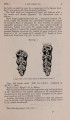
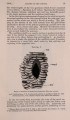
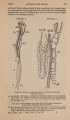
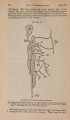
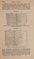
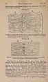
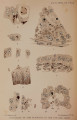
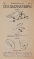
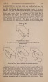
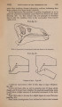
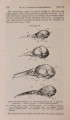
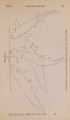
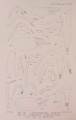
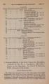
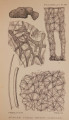
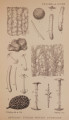
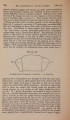
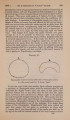
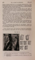
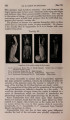
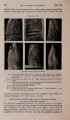
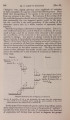
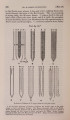
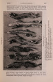
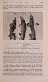
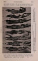
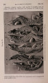
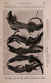
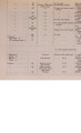
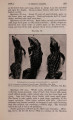
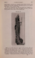
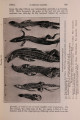
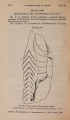
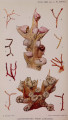
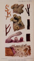
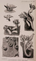
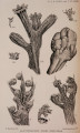
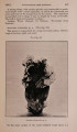
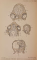
Show 80 DR. J. W. JENKINSON ON THE [Feb. 6, (/3) In addition to these typical cells I have found in the Cow elements which look remarkably like goblet-cells. They are to be seen (PI. III. fig. 8, gl.) wedged in between the ordinary cubical cells which cover the villi. Each cell contains a goblet-cavity filled with a granular coagulum; the nucleus is small and pressed against the side of the goblet. I have not succeeded in getting the granules to stain with muci-carmine or muchammtein. (•y) The large oval binucleate cells (PI. III. fig. 8, bi.), found in both the extra-cotyledonary trophoblast and upon the villi, have been described by both Bonnet and Kolster. Erch cell has a very definite superficial membrane, a dense finely granular cytoplasm, and two large oval nuclei provided with a rich reticulum of coarse chromatin granules and two or more plasmosomes. The nuclei may divide mitotically. According to Kolster, these cells are maternal leucocytes which have migrated through the uterine epithelium, grown at the expense of the cell-debris accumulated in the lumen uteri, doubled their nuclei by amitotic division, and become incorporated in the trophoblast. It is true that these or closely similar cells are occasionally found free between the foetal and maternal tissues; but apart from that I do not believe there is the least evidence for the view put forward by Kolster. That leucocytes migrate in large numbers during gestation through the maternal epithelium and are found in the " uterine milk " is certainly an indisputable fact; these cells are, however, far larger than any leucocytes that I have ever found, and quite dissimilar to any that I a m acquainted with. Moreover, Kolster does not figure a good series of the alleged intermediate stages between the unmodified white corpuscles and these very peculiar cells. The question must remain an open one until the mode of first appearance of these elements in the unattached blastocyst has been ascertained *. 3. The u Diverticula Allanto id is." The ends of the chorionic sac-placed in the cornua uteri-are produced into long, tapering filaments, supposed by the earlier embryologists to be diverticula of the allantois pushed through perforations in the chorion. Bonnet has shown (for the Sheep) * Assheton has now shown that these cells are of foetal origin. See postscript to this paper. Explanation of Text-fig. 29 (opposite). Sheep.-Formation of new crypt-cavities by the downgrowth of cell-masses from patches of unmodified-not flattened-epithelium. In fig. a the solid downgrowth is shown in continuity with the crypt epithelium ; in fig. b it is cut across; in fig.e it has a lumen communicating with the crypt-cavity above; in fig. d the lumen though well-developed is not yet open ; and in fig. c the lumen is as yet exceedingly small. All the figures taken from the basal crypts of a cotyledon of a foetus measuring 14 cm. (3rd month according to Kolster.) |