| OCR Text |
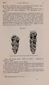
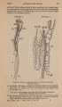
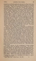
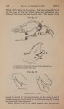
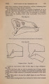
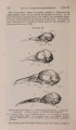
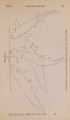
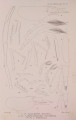
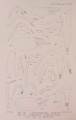
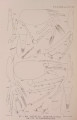
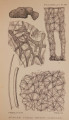
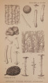
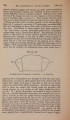
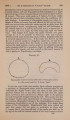
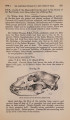
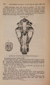
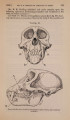
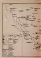
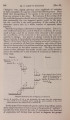
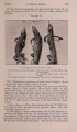
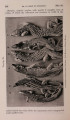
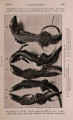
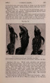
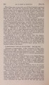
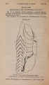
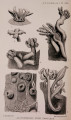
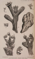
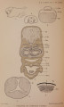
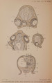
Show 1906.] CYCLOPIA IN OSSEOUS FISHES. 445 increased as also has the lens. The lens-cavity is not completely occupied by fibres, a space being left anteriorly which is filled by small round cells. Retina, choroid, cornea, vitreous humour, and sclerotic are well developed. The single choroidal fissure leads back to a large optic nerve formed, as above stated, by the union of the two optic tracts (fig. 4). There are two choroidal glands, one on either side of the optic pore. They are supplied, as usual, by choroidal arteries coming from the pseudobranchs. The following eye-muscles are present:-two superior obliques, arising from the supra-orbital bars; two superior recti, arising along with two inferior recti from the fibrous capsule of the brain in front of the hypophysis; two external recti, which are normal in origin and are inserted into the right and left sides respectively of the eyeball. The inferior recti are united close to their insertion into the eyeball. Inferior obliqui and internal recti are absent. (B) Cyclopia with Fusion of Structures in the Mid-brain and of the Cerebrcd Lobes. Three of my specimens exhibit this condition, two of them possessing a single median eye, while the third, although showing the other essential features of cyclopia, has a pair of small closely-approximated eyes. 1. The specimen which has a single eye resembles type A in general appearance, except as regards its mouth-parts. In place of the lower jaw there is a membranous flap on either side projecting downwards and forwards from below the eye. In place of the lower jaw arcade there is a narrow mesial process projecting forwards to end just between the flaps. Microscopic examination of the flaps shows that they contain externally a number of young teeth and internally a commencing membranous ossification. They are probably to be compared with ununited maxillary processes, and in this respect they resemble the hornlike structures found by Paolucci* in his cyclopean Skate. The mesial process above mentioned contains a much elongated symphysis of the lower jaw, the Meckel's bars of which diverge little from one another and articulate with suspensoria which are similarly approximated. Skeleton.-The trabecular cranii are represented by a single exceedingly short bar projecting downwards and forwards towards the wall of the pharynx. Quite separate from this are the palato-quadrates, the anterior ends of which, uniting below the eye, form a mesial plate replacing the defective trabecular The supraorbitals are different in the two specimens: in one they unite anteriorly in the frontal process, giving rise to a small olfactory capsule ; in the other they are short and extend no further forward than the middle of the fore-brain. In this latter case * Atti dclla Societa Italiana di Scienzc Naturali, vol. xvii. 1874. |