| OCR Text |
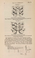
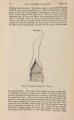
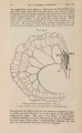
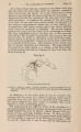
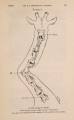
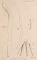
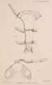
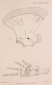
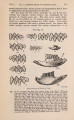
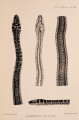
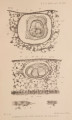
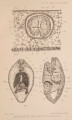
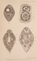
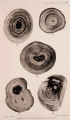
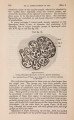
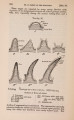
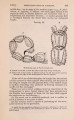
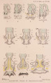
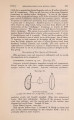
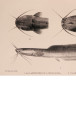
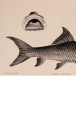
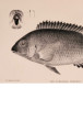
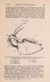
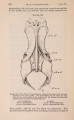
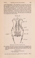
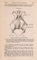
Show 1902.] ORIGIN OF PEARLS. 149 tissue fibrils (PI. XIV. fig. 2), but soon the tissues of the host give rise to an epithelial layer, which lines the space and ultimately becomes the pearl-sac (PL X V . fig. 5, s.). This epithelium appears to arise quite independently of the outer epidermis, and is no doubt due to a specific stimulation on the part of the parasite, as other parasites, e. g. Sporocysts, Cestode larvae, &c, are not surrounded by such a sac. At first a few cells appear (PI. X I V . figs. 2, 3, pr.), which proliferate and arrange themselves along the walls of the cavity. These cells are larger than the connective-tissue corpuscles, and more susceptible to stains. They are flattened and polygonal in surface view. Their nuclei (PI. X I V . fig. 3, n.) are large and spherical, and show the conspicuous chromatin reticulum and distinct nucleolus that characterize the nuclei of embryonic or rapidly dividing tissues. I have not been able to find the nuclei of these cells actually undergoing division. The proliferating sheet of cells ultimately surrounds the parasite and becomes the sac. From the first these cells are basally continuous with fibres of connective tissue (PI. X I V . fig. 3, c.t.). Their transformation into the pearl-sac is a gradual one, and every step can be traced in sections of the parasites in situ,. If the Trematode larva completes its maximum possible term of life it dies, and the tissues of the body break down to form a structureless mass, which retains the form of the parasite owing to the rigid cuticle. In this mass arise one or more centres of calcification (PI. X V I . fig. 8), and the precipitation of carbonate of lime goes on until the whole larva is converted into a nodule which has the calco-sphaeritic structure already described for the nucleus. The granular matter surrounding the worm, if present, also undergoes calcification. The epithelium of the sac then begins to shed a cuticle of 1 conchyolin (PI. X I V . fig. 1), and from this point the growth of the pearl probably takes place on the same lines and at the same rate as the thickening of the shell. The sac sometimes begins to form pearly substance before the worm is completely calcified (PI. XVII. fig. 16). The Distomid larvae sometimes leave the sac formed around them, and voluntarily migrate into other parts of the body before again settling down. Empty sacs may be found in the mantle, and old specimens of the larva (distinguishable from recently immigrated ones by their darker colour and laden excretory organs) sometimes occur free between the mantle and the shell. The occurrence of pearls in which the nucleus is not a Trematode but merely a few refractive granules (PI. XVII. fig. 13) can be accounted for in this manner. Some compound pearls are evidently formed by short migrations on the part of the Cercariae, which leave a small amount of |