| OCR Text |
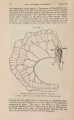
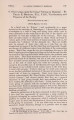
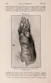
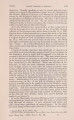
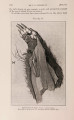
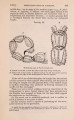
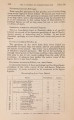
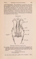
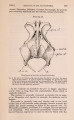
Show 1902.] ADAPTATIONS IN DIPROTODONT MARSUPIALS. 21 of food-material, and the mechanical pressure of this lieavy load might have produced expansions, which, if localized, formed bags or sacculi. The first part of the colon might be expected to have been strongly affected by this pressure. It is consequently natural that some wide sacculi should be formed there, and it is these distensions which have been described and figured by the authors as caecum, although they are derived purely from the colon. It is also easy to understand that when this distension took place the originally transverse caeco-colic plica was drawn or turned out of place to its present longitudinally-running direction. That the mechanical pressure of the contents of the colon has really played important parts in transforming it to its present shape, may also be proved by another fact. By a broad band opposite the mesentery the colon is sacculated, which, of course, is also an adaptation to its function. At the place where the colon is most closely, by a very short mesentery, soldered to the back of the abdominal cavity, the pressure of the contents would, thanks to this fixation, be more effective. There has thus been formed two large sacculi, which give the colon at that place a size amounting to twice that of its usual width. The shape and size of these sacculi are identical in two specimens which I have seen. This confirms the correctness of the statement; and I think it is these which Owen means when he says: " One of these sacculi was so much longer than the rest as to almost merit special notice as a second caecum." Peyer's patches of comparatively large size, 1 to 2 cm. in diameter, are scattered in considerable numbers in the walls of the colon, especially in its middle parts. The material which I have used for this study has long been preserved in spirit, and the measurements are perhaps therefore not so much to be relied upon. It may, however, be mentioned that the small intestine measured in one specimen about 410 cm., the caecal rudiment 6 cm. from its blind end to its opening, and the large intestine 840 cm. The " secondary caecum " is situated nearly at the middle, or 430 cm. from the end. Even if these measurements are imperfect in the detailed statements, they show satisfactorily that the large intestine has been strongly developed. Probably it is fully eight times the length of the animal, or even more than in the Koala. The interior surface of the duodenum in Phalanger shows very plainly a reticulate structure, larger primary and smaller secondary plicae may easily be distinguished. It offers thus some faint resemblance in appearance to the structure of the reticulum of a ruminant. The plicae are in both cases formed by coalescence of papillae. The villi of the intestine are well developed on the ridges forming the network, but some are also scattered in the interspaces. Lower down the small intestine this reticulate structure is less conspicuous, but m y material is not in such good condition that I can say where it entirely disappears. The jejunum appears, however, quite smooth. |