| OCR Text |
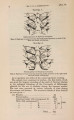
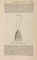
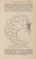
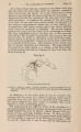
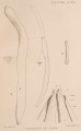
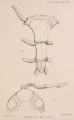
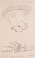
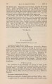
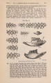
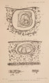
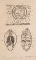
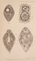
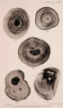
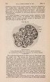
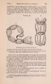
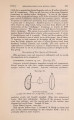
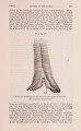
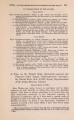
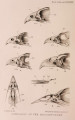
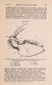
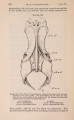
Show 148 DR. II. LYSTER JAMESON ON THE [Mar. 4, pearl are, like the shell, formed by the calcification of the cuticle of the living cells, and owe their structure to the special characters of that membrane or of the underlying epidermis. A section of a decalcified pearl shows the nucleus, in which the cuticle and sometimes the suckers of the Distomum can be distinguished. Occasionally the outlines of the soft parts (e. g., pharynx and digestive caeca) are still visible, as in PI. X I V . fig. 1, ph. &, dig. More generally, however, nothing can be seen but a mass of yellowish-brown granular substance surrounded by the cuticle (text-fig. 22). There is often a certain amount of refractive granular matter associated with the remains of the worm, probably an excretion; and, if the parasite migrates out of the sac, this may form the inconspicuous nucleus of a pearl. Just as the peripheral parts of a pearl present, when ground down to a thin section, a similar structure to that of the shell, so the conchyolin basis of a decalcified pearl shows the same characters. The outermost layer of the latter is uncalcified and continuous with the cuticle of the cells of the sac, just as the outer mantle-epidermis is attached to the inner surface of the shell (PI. X I V . fig. 1, con.1). There is no organic union between the conchyolin and the nucleus. The sac containing the pearl is composed of a simple columnar epithelium (PI. X I V . fig. 1 & text-fig. 22, s.), which in its histological structure, as well as in its power of secreting as a cuticle the conchyolin basis of the pearl, is indistinguishable from the outer epidermis of the mantle. Blood-spaces, containing corpuscles, are well developed around the sac. Such a pearl cannot then be compared-as some writers have suggested-with the concretions or calculi of cholesterin or other substances found in the vertebrate body, but rather with the structures sometimes found in epidermoid tumours and atheroma cysts. Origin and Development of the Pearl. The Trematode enters Mytilus edulis as a tailless Cercaria, and at first m ay often be found between the mantle and shell. It is probable that it reaches this position by boring through the mantle, but I have not yet been able to find one in the act of doing so. The larvae creep about on the inner surface of the shell, and, after a while, again enter the connective tissue of the mantle, where they come to rest, assuming a spherical form. They seem to avoid the more muscular parts of the mantle-no doubt because the absence of a definite boring apparatus makes it difficult for them to pass through the latter. W h e n embedded in the tissues they are visible to the naked eye as little yellowish spots, about | m m . in diameter. At first the worm only occupies a space lined by connective- |