| OCR Text |
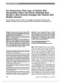
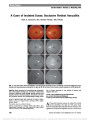
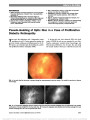
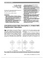
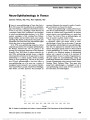
Show The Retinal Nerve Fiber Layer of Patients With Neuromyelitis Optica and Chronic Relapsing Optic Neuritis is More Severely Damaged than Patients With Multiple Sclerosis Denis B. Bichuetti, MD, PhD, André S. de Camargo, MD, Alessandra B. Falcão, MD, Fabiana F. Gonçalves, MD, Ivan M. Tavares, MD, PhD, Enedina M.L. de Oliveira, MD, PhD Backgroud: To compare the retinal nerve fiber layer (RNFL) in eyes of patients with relapsing remitting multiple sclero-sis (RRMS), neuromyelitis optica (NMO) and chronic relaps-ing inflammatory optic neuritis (CRION). Methods: Evaluation of 62 patients with RRMS, NMO, and CRION in a cross-sectional study with spectral domain optical coherence tomography. Results: A total of 124 eyes were evaluated (96 RRMS, 18 NMO, and 10 CRION). Frequency of optic neuritis for each disease was: 34% for RRMS, 84% for NMO, and 100% for CRION. Visual acuity and RNFL thickness were significantly worse in NMO and CRION eyes than in RRMS, but there were no differences between NMO and CRION eyes. A RNFL of 41 mm was 100% specific for optic neuritis associated with NMO and CRION when compared to RRMS. Conclusion: This study established RNFL values to differ-entiate optic neuritis of RRMS from NMO and CRION. Although similarities observed between NMO and CRION eyes might suggest that they are within the same disease spectrum, it is still recommended that these 2 conditions be differentiated on clinical grounds. Optical coherence tomography serves as an additional diagnostic tool and can be used to monitor disease progression. Journal of Neuro-Ophthalmology 2013;33:220-224 doi: 10.1097/WNO.0b013e31829f39f1 © 2013 by North American Neuro-Ophthalmology Society The differential diagnosis of optic neuritis (ON) con-tinues to expand. The clinician must not only differ-entiate ON associated with multiple sclerosis, (1) infectious causes, and autoimmune conditions (2,3) but must also include neuromyelitis optica (NMO) (4) and cases of recurrent ON without clinical evidence of disease beyond the anterior visual pathways (5-10). The discovery of aquaporin-4 antibody (NMO-IgG) (11) has shown that many inflammatory diseases of the central nervous system, including recurrent longitudinally extensive transverse myelitis, recurrent ON, and recurrent brainstem and encephalic syndromes, may fall within the disease spec-trum of aquaporin-4 autoimmunity called NMO spec-trum disorders (NMO-SD) (12,13). In their original report of chronic relapsing inflammatory optic neuritis (CRION), Kidd et al (5) described patients with bilateral inflammatory optic neuritis with recurrent relapses over time and that worsened upon steroid or immu-nosuppression withdrawal. This led to reports differentiat-ing other forms of relapsing ON from CRION (6-8). It is unclear whether there is a chronic progressive or relapsing course in CRION patients, because these patients tend to relapse upon medication withdrawal in weeks to months. Departamento de Neurologia e Neurocirurgia (DBB, ABF, EMLdO) and Departamento de Oftalmologia (ASdC, FFG, IMT), Universidade Federal de São Paulo, São Paulo, Brazil. D.B. Bichuetti has received speaking honoraria from Bayer Health Care, TEVA, and Merck Serono and travel expenses to scientific meetings from Bayer Health Care, Merck Serono, Novartis, and Teva. E.M.L. de Oliveira received compensations for participating in meetings sponsored by Bayer Health Care, Biogen Idec, Merck Se-rono, Novartis, and Teva. A.B. Falcão received travel expenses to scientific meetings from Bayer Health Care, Merck Serono, and Novartis. I.M. Tavares has received travel expenses to scientific meetings from Alcon, Allergan, and Merck Sharp & Dohme and is a medical advisor at Allergan and Merck Sharp & Dohme. A.S. de Camargo reports no conflicts of interest. Denis B. Bichuetti had full access to all of the data in the study and takes responsibility for the integrity of the data and the accuracy of the data analysis. Supplemental digital content is available for this article. Direct URL citations appear in the printed text and are provided in the full text and PDF versions of this article on the journal's Web site (www. jneuro-ophthalmology.com). Address correspondence to Denis B. Bichuetti, MD, PhD, Disciplina de Neurologia, Universidade Federal de São Paulo, (Neurology Discipline, Federal University of São Paulo), Rua Botucatu, 740, 04023-900 São Paulo, SP, Brazil; E-mail: bichuetti@unifesp.com 220 Bichuetti et al: J Neuro-Ophthalmol 2013; 33: 220-224 Original Contribution Copyright © North American Neuro-Ophthalmology Society. Unauthorized reproduction of this article is prohibited. Possibly, some patients with CRION would fit into NMO-SD (14,15). Optical coherence tomography (OCT) has been used to evaluate RNFL thickness in various diseases, including multiple sclerosis, neuromyelitis optica, and clinical isolated syndromes (16-19). We performed a single center cross-sec-tional study comparing the visual acuity and RNFL thickness with spectral domain optical coherence tomography (SD-OCT) in patients with relapsing remitting multiple sclerosis (RRMS), NMO, and CRION, to assess whether this tech-nique could distinguish ON in these 3 disorders. METHODS We recruited patients with RRMS, NMO, and CRION seen in 2010 and 2011 in the neuroimmunology clinic at the Federal University of São Paulo, São Paulo, Brazil. Patients had to satisfy diagnostic criteria for RRMS (20) and NMO (21), while those with CRION experienced at least 2 inflammatory ON episodes 30 days apart without clinical or radiological signs of disease activity elsewhere in the central nervous system. We classified the patients with CRION if they had suffered .1 relapse of ON in the same, fellow, or both eyes, regardless of whether they were previously immunosuppressed. All patients had at least 1 brain and spinal cord magnetic resonance imag-ing (MRI) study at the first evaluation and were screened for autoimmune and systemic causes. This included testing for human immunodeficiency virus, hepatitis B and C, human T-cell lymphotropic virus, syphilis, toxoplasmosis, and cyto-megalovirus. A thorough review of systems was completed on CRION patients to exclude a potential toxic or genetic cause. Patients had an expanded disability status scale (EDSS) score of 0-5.5. Peripapillary RNFL thickness was measured by SD-OCT, using the Spectralis software (version 4.0, Heidelberg Engineering, Dossenheim, Germany). Only well-focused, evenly illuminated, and centered images were included in the analysis. We collected demographic, clinical, laboratory, and MRI data of all patients. NMO-IgG testing using indirect immunofluorescence was performed in all patients with NMO or CRION seen after 2007, as this test was not available in Brazil before that date. Thus, some samples came from patients already on immunosuppressive medication, which is known to reduce the sensitivity of the test (22,23). To avoid bias because of disease duration, we normalized the EDSS (24) on last follow-up visit and total number of relapses by the total time of disease (in years), thus using the pro-gression index (PI) and annualized relapse rate (25). Because patients with CRION would have a maximum EDSS of 4 due to conversion of visual functional system scores on the final EDSS step, we categorized each patient's visual acuity (VA) in each eye according to the EDSS visual functional system score (0 = normal; 1 = VA 20/20 to 20/29; 2 = VA 20/30 to 20/59; 3 = VA 20/60 to 20/99; 4 = 20/100 to 20/199; and 5 = VA worse than 20/200) and calculated the PI exclusively for this system. Almost all patients received treatment with 1 or a com-bination of drugs including corticosteroids, azathioprine, methotrexate, cyclophosphamide, interferon beta, glatiramer acetate, natalizumab, and IV immunoglobulin. Statistical analysis was performed using GraphPad Prism version 5.0f, with data presented as mean ± SD or median and interquartile range. Unpaired t test was used when comparing 2 groups, and analysis of variance was used when appropriate. A receiver-operator characteristic curve (ROC curve) was established to estimate sensitivity and sensitivity to differentiate these diseases through the use of OCT. The Internal Review Board of the Universidade Federal de São Paulo approved our study, and written informed consent was obtained from all subjects before the performance of the neurophtalmologic and SD-OCT examinations. RESULTS Sixty-two patients completed neuroophthalmologic and SD-OCT examinations: 48 with RRMS, 9 with NMO TABLE 1. OCT, EDSS, and PI results in patients with optic neuritis Disease Age of Onset Disease Duration, y History of Optic Neuritis (Eyes) Age OCT Was Performed RNFL, mm EDSS Score RRMS, n = 48 29.9 (±10.4) 8.7 (±5.9) 33 (34%) 38.6 (±11.8) 87.7 (±15.3) 2.2 (±1.1) NMO, n = 9 30.3 (±9.3) 8.6 (±3.2) 15 (±84%) 39.0 (±10.5) 65.8 (±20.9) 3.3 (±0.8) CRION, n = 5 36.2 (±6.7) 7.0 (±8.9) 10 (100%) 43.0 (±6.3) 48.9 (±15.3) 3.6 (±0.9) Disease EDDS FS Score Visual Pyramidal Cerebellar Sensory Brainstem PI PI Visual RRMS, n = 48 1.2 (±1.5) 1.3 (±1.1) 0.4 (±0.9) 0.7 (±0.9) 0.1 (±0.3) 0.3 (±0.2) 0.2 (±0.3) NMO, n = 9 3.5 (±1.8) 1.1 (±0.6) 0.0 (±0.0) 0.7 (±1.0) 0.0 (±0.0) 0.4 (±0.2) 0.4 (±0.3) CRION, n = 5 4.2 (±1.2) 0.0 (±0.0) 0.0 (±0.0) 0.0 (±0.0) 0.0 (±0.0) 1.0 (±0.6) 1.1 (±0.7) Parentheses denote standard deviation. CRION, chronic relapsing inflammatory optic neuritis; EDSS, expanded disability status score; FS, functional system; NMO, neuromyelitis optica; OCT, optical coherence tomography; PI, progression index; RNFL, retinal nerve fiber layer; RRMS, relapsing remitting multiple sclerosis. Bichuetti et al: J Neuro-Ophthalmol 2013; 33: 220-224 221 Original Contribution Copyright © North American Neuro-Ophthalmology Society. Unauthorized reproduction of this article is prohibited. (8 relapsing and 1 monophasic), and 5 with CRION. Of the 124 eyes evaluated with SD-OCT, only 99 were of sufficient quality for interpretation (79 from RRMS, 12 from NMO, and 8 from CRION) (See Supplemental Digital Content, Table 1, http://links.lww.com/WNO/A80). The main reasons for inadequate quality were nystagmus and difficulty in maintaining steady fixation because of impaired VA. Thirty-four percent of eyes from patients with RRMS, 84% of the eyes from patients with NMO, and all eyes from patients with CRION experienced at least 1 episode of ON. Age of onset and age at SD-OCT measurements did not differ between the groups (ANOVA with Bonferroni multiple comparison test) and disease duration was also similar (ANOVA with Dunn multiples comparison test) (Table 1). Last measured VA was worse in NMO and CRION eyes than RRMS (P , 0.0001 for NMO or CRION vs RRMS and P = 0.4703 for NMO vs CRION), but there was no difference between NMO and CRION (P = 0.4703). The PI for visual functional system of patients with NMO was worse than patients with RRMS (P , 0.0001), and patients with CRION had higher rates than patients with NMO (P , 0.0075). Clinical comparison of other aspects of the expanded EDSS was not possible, as patients with NMO and CRION presented with fewer neurologic symptoms, and the analysis would be biased towards worse results in RRMS patients. Since only patients with mild-to-moderate EDSS were recruited, patients with RRMS pre-sented low scores on other functional systems as well. NMO-IgG testing was performed in all patients categorized as NMO or CRION, with 45% and 20% positivity, respec-tively. Patients who fulfilled criteria for multiple sclerosis were not routinely tested for NMO-lgG, because this test has limited value in the typical multiple sclerosis patient (26). However, 7 patients with RRMS were tested for NMO-IgG, all with negative results. In evaluating eyes with ON, patients with RRMS had RNFL substantially thicker than patients with NMO or CRION (Table 2). There was no statistical difference in the RNFL between NMO and CRION eyes. We analyzed eyes by the number of ON episodes (0, 1, 2, and $3) to identify whether the differences observed in RNFL were an effect of the number of relapses or severity (Tables 3 and 4). This appeared to have no effect, that is, NMO and CRION eyes were more severely affected than RRMS, but without a signif-icant difference between them (Table 4). Although the number of eyes was small, there was no difference in RNFL thickness and VA between the 63 eyes from RRMS patients and the 3 eyes from NMO patients who lacked a history of ON. An ROC curve was created comparing eyes that had ON from patients with RRMS, NMO, and CRION to establish sensitivity and specificity values that could differentiate these conditions. The eyes from RRMS patients were used as controls and the eyes from NMO and NMO + CRION were pooled together (Fig. 1). The comparison of RRMS eyes to NMO with an ROC curve presented an area under the curve (AUC) of 0.7991 with P = 0.0083, and when using NMO and CRION eyes pooled together as patients and RRMS as controls, an AUC of 0.8507 with P = 0.0001. In both cases, a RNFL thickness of 41 mm or less was 100% specific for ON associated with NMO or CRION when compared to RRMS. None of the eyes from RRMS patients had values below 41 mm, as opposed to 2 with NMO (1 patient) and 4 with CRION (3 patients). DISCUSSION The recognition of distinct clinical and pathophysiological features of NMO-SD has placed these conditions into TABLE 2. Retinal nerve fiber layer thickness comparisons in eyes with optic neuritis P RRMS vs NMO 0.0040 RRMS vs CRION ,0.0001 NMO vs CRION 0.2149 Unpaired t test was used in analysis. CRION, chronic relapsing inflammatory optic neuritis; NMO, neuromyelitis optica; RRMS, relapsing remitting multiple sclerosis. TABLE 3. Comparison of EDSS scores and RNFL thickness in patients with RRMS, NMO, and CRION based on the number of episodes of optic neuritis Episodes RRMS NMO CRION n Visual FS RNFL n Visual FS RNFL n Visual FS RNFL 0 63 0.0 (0.0-1.5) 92.4 (±14.0) 3 0.0 (0.0-2.0) 85.7 (±20.5) 0 NA* NA* 1 26 2.0 (0.75-4.3) 80.1 (±14.8) 8 5.0 (2.0-5.0) 65.3 (±18.3) 5 5.0 (3.5-5.0) 50.6 (±17.1) 2 4 2.0 (1.3-2.0) 61.7 (±7.1) 7 5.0 (3.0-5.0) 47.0 (±6.1) 2 4.1 (4.0-4.1) 37.5 (±0.7) $3 3 1.0 (0.0-3.0) 81.3 (±17.5) 0 NA* NA* 3 5.0 (2.0-5.0) 63.0† Data is represented as median and 25th and 75th percentiles in parenthesis. *No eye in this category. †Single eye. CRION, chronic relapsing inflammatory optic neuritis; EDSS, expanded disability status score; FS, functional system; NMO, neuromyelitis optica; RNFL, retinal nerve fiber layer; RRMS, relapsing remitting multiple sclerosis. 222 Bichuetti et al: J Neuro-Ophthalmol 2013; 33: 220-224 Original Contribution Copyright © North American Neuro-Ophthalmology Society. Unauthorized reproduction of this article is prohibited. a separate category of CNS inflammatory disorders (12,27,28). In this context, we demonstrated that changes in RNFL thickness of optic neuritis patients with CRION are similar to NMO, and both are distinct from RRMS. Previous reports of relapsing ON not related to RRMS have included patients with severe visual loss and a mean age of onset between 20 and 50 years. Nearly 50% of relapses occur within 1 year, but in some up to 10 years (5-8, 29, 30). The rate of NMO-IgG positivity in relapsing ON has been reported between 20% and 50% (7, 8, 10, 29, 30), and with nearly 50% of patients with relapsing ON converting to NMO (29,31). We recognize the limitations of our study. All patients with RRMS received immunomodulatory treatment, and all patients with NMO and all but 1 with CRION were given immunosuppressive medication. This likely biased our analysis due to treatment duration and intensity. Patients exclusively with ON relapses were less likely to receive treatment or receive it later in the clinical course than those with additional neurologic findings (data not shown). This delay could have contributed to differences in RNFL thickness between NMO and CRION. Also, the selection of RRMS and NMO patients with low-to-moderate EDSS scores created a cohort bias. Yet, this allowed us to include patients within the first 10 years of disease onset, producing a more homogenous sample of patients with similar disease duration. Although statistical comparison of RNFL thick-ness from patients with RRMS and NMO with 1 or 2 ON episodes was not significant (Table 4), differing from the analysis of all eyes together (Table 2), we believe that this trend was due to splitting the groups into smaller samples and reducing statistical power. The inclusion of patients after their first clinical event, as performed by Collongues et al (32), requires a longitudinal study, which is currently underway in our center. Our study has shown that in patients with ON, measurement of RNFL thickness is similar in NMO and CRION patients compared to those with RRMS. Although the value of 41 mm for RNFL thickness is 100% specific for NMO and CRION, and may assist in differentiation from TABLE 4. Statistical analysis of EDSS visual system function score and RNFL thickness in patients with RRMS, NMO, and CRION based on the number of episodes of optic neuritis Category RRMS vs NMO RRMS vs CRION NMO vs CRION Visual FS Episodes of ON 0 0.6501 * * 1 0.0671 0.0259 0.4949 2 0.0042 0.0036 0.8074 $3 † 0.1161 † RNFL Episodes of ON 0 0.4355 * * 1 0.0532 0.0008 0.2037 2 0.0531 0.0198 0.1280 $3 † ‡ †,‡ Unpaired t test was used in all analysis. *No eye with CRION in this category. †No eye with NMO in this category. ‡Comparison not possible as there is only 1 eye with CRION with adequate quality optical coherence tomography in this category. CRION, chronic relapsing inflammatory optic neuritis; EDSS, expanded disability status score; FS, functional system EDDS score; NMO, neuromyelitis optica; ON, optic neuritis; RNFL, retinal nerve fiber layer; RRMS, relapsing remitting multiple sclerosis. FIG. 1. Receiver-operator characteristic curves for RNFL thickness in optic neuritis patients with RRMS, NMO, and CRION. Left: Curve built using RRMS as controls and NMO + CRION pooled together as subjects area under the curve (AUC) = 0.8507, P = 0.0001. Right: Curve built using RRMS as controls and NMO as subjects. AUC = 0.7991, P = 0.0083. CRION, chronic relapsing inflammatory optic neuritis; NMO, neuromyelitis optica; RNFL, retinal nerve fiber layer; RRMS, relapsing remitting multiple sclerosis. Bichuetti et al: J Neuro-Ophthalmol 2013; 33: 220-224 223 Original Contribution Copyright © North American Neuro-Ophthalmology Society. Unauthorized reproduction of this article is prohibited. RRMS, establishing the diagnosis of these 3 disorders still rests on clinical, neuroimaging, and serologic findings. REFERENCES 1. Hickman SJ, Dalton CM, Miller DH, Plant GT. Management of acute optic neuritis. Lancet. 2002;360:1953-1962. 2. Kupersmith MJ, Burde RM, Warren FA, Klingele TG, Frohman LP, Mitnick H. Autoimmune optic neuropathy: evaluation and treatment. J Neurol Neurosurg Psychiatry. 1988;51:1381-1386. 3. Gelwan MJ, Kellen RI, Burde RM, Kupersmith MJ. Sarcoidosis of the anterior visual pathway: successes and failures. J Neurol Neurosurg Psychiatry. 1988;51:1473-1480. 4. Papais-Alvarenga RM, Carellos SC, Alvarenga MP, Holander C, Bichara RP, Thuler LC. Clinical course of optic neuritis in patients with relapsing neuromyelitis optica. Arch Ophthalmol. 2008;126:12-16. 5. Kidd D, Burton B, Plant GT, Graham EM. Chronic relapsing inflammatory optic neuropathy (CRION). Brain. 2003;126:276-284. 6. Pirko I, Blauwet LK, Lesnick TG, Weinshenker BG. The natural history of recurrent optic neuritis. Arch Neurol. 2004;61:1401-1405. 7. Jarius S, Frederikson J, Waters P, Paul F, Akman-Demir G, Marignier R, Franciotta D, Ruprecht K, Kuenz B, Rommer P, Kristoferitsch W, Wildemann B, Vincent A. Frequency and prognostic impact of antibodies to aquaporin-4 in patients with optic neuritis. J Neurol Sci. 2010;298:158-162. 8. Petzold A, Pittock S, Lennon V, Maggiore C, Weinshenker BG, Plant GT. Neuromyelitis optica-lgG (aquaporin-4) autoantibodies in immune mediated optic neuritis. J Neurol Neurosurg Psychiatry. 2010;81:109-111. 9. Kim SH, Kim W, Li XF, Jung IJ, Kim JH. Clinical spectrum of CNS aquaporin-4 autoimmunity. Neurology. 2012;78:1179-1185. 10. Falcão AB, Oliveira EML, Bichuetti DB, Camargo AS, Tavares IM, Souza NA, Fragomeni MO, Gabbai AA. Chronic relapsing inflammatory optic neuropathy: a Brazilian Cohort. Mult Scler. 2011;17:S58. 11. Lennon VA, Wingerchuk DM, Kryzer TJ, Pittock SJ, Lucchinetti CF, Fujihara K, Nakashima I, Weinshenker BG. A serum autoantibody marker of neuromyelitis optica:distinction from multiple sclerosis. Lancet. 2004;364:2106-2112. 12. Jarius S, Ruprecht K, Wildemann B, Keumpfel T, Ringelstein M, Geis C, Kleiter I, Kleinschnitz C, Berthele A, Brettschneider J, Hellwig K, Hemmer B, Linker RA, Lauda F, Mayer CA, Tumani H, Melms A, Trebst C, Stangel M, Marziniak M, Hoffmann F, Schippling S, Faiss JH, Neuhaus O, Ettrich B, Zentner C, Guthke K, Hofstandt-van Oy U, Reuss R, Pellkofer H, Ziemann U, Kern P, Wandinger KP, Bergh FT, Boettcher T, Langel S, Liebetrau M, Rommer PS, Niehaus S, Munch C, Winkelmann A, Zettl UU, Metz I, Veauthier C, Sieb JP, Wilke C, Hartung HP, Aktas O, Paul F. Contrasting disease patterns in seropositive and seronegative neuromyelitis optica: a multicenter study of 175 patients. J Neuroinflam. 2012;9:14. 13. Matiello M, Jacob A, Wingerchuk DM, Weinshenker BG. Neuromyelitis optica. Curr Opin Neurol. 2007;20:255-260. 14. Jacob A, McKeon A, Nakashima I, Sato DK, Elsone L, Fujihara K, de Seze J. Current concept of neuromyelitis optica (NMO) and NMO, spectrum disorders. J Neurol Neurosurg Psychiatry. 2012 Nov 10 (epub ahead of print). 15. De Seze J. Atypical forms of optic neuritis. Rev Neurol (Paris). 2012;168:697-701. 16. Monteiro ML, Fernandes DB, Apostolos-Pereira SL, Callegaro D. Quantification of retinal nenural loss in patients with neuromyelitis optica and multiple sclerosis with or without optic neuritis using fourier-domain optical coherence tomography. Invest Ophthalmol Vis Sci. 2012;53:3959-3966. 17. Morrow MJ, Wingerchuk D. Neuromyelitis optica. J Neuroophthalmol. 2012;32:154-166. 18. Watson GM, Keltner JL, Chin EK, Harvey D, Nguyen A, Park SS. Comparison of retinal nerve fiber layer and central macular thickness measurements among five different optical coherence tomography instruments in patients with multiple sclerosis and optic neuritis. J Neuroophthalmol. 2011;31:110-116. 19. Trip SA, Schlottmann PG, Jones SJ, Kallis C, Altmann DR, Garway-Heath DF, Thompson AJ, Plant GT, Miller DH. Scanning laser polarimetry quantification of retinal nerve fiber layer thinning following optic neuritis. J Neuroophthalmol. 2010;30:234-242. 20. Polman CH, Reingold SC, Banwell B, Clanet M, Cohen JA, Flippi M, Fujiharak, Havrdova A, Hutchinson M, Kappos L, Lublin FD, Montalban X, O'Connor P, Sandberg-Wolheim M, Thompson AJ, Waubant E, Weinshenker B, Wolinsky JS. Diagnostic criteria for multiple sclerosis:2010 revisions to the McDonald criteria. Ann Neurol. 2011;69:292-302. 21. Wingerchuk DM, Lennon VA, Luchinetti CF, Pittock SJ, Weinshenker BG. The spectrum of neuromyelitis optica. Lancet Neurol. 2007;6:805-815. 22. Weinstock-guttman B, Miller C, Yeh E, Stosic M, Umhauer M, Batra N, Munschauer F, Zivadinov R, Ramanathan M. Neuromyelitis optica immunoglobulins as a marker of disease activity and response to therapy in patients with neuromyelitis optica. Mult Scler. 2008;14:1061-1067. 23. Jarius S, Aboul-Enein F, Waters P, Kuenz B, Hauser A, Berger T, Lang W, Reindl M, Vincent A, Kristoferitsch W. Antibody to aquaporin-4 in the long-term course of neuromyelitis optica. Brain. 2008;131:3072-3080. 24. Kurtzke JF. Rating neurologic impairment in multiple sclerosis; an expanded disability status scale (EDSS). Neurology. 1983;33:1444-1452. 25. Weinshenker BG, Bass B, Rice GP, Noseworthy J, Carriere W, Baskerville J, Ebers course and disability. Brain. 1989;112:133-146. 26. Rubiera M, Rio J, Tintore M, Nos C, Rovira A, Tellez N, Montalban X. Neuromyelitis optica diagnosis in clinically isolated syndromes suggestive of multiple sclerosis. Neurology. 2006;66:1568-1570. 27. Bizzoco E, Lolli F, Repice AM, Hakiki B, Falcini M, Barilaro A, Taiuti R, Siracusa G, Amato MP, Biagioli T, Lori S, Moretti M, Vinattieri A, Nencini P, Massacesi L, Mata S. Prevalence of neuromyelitis optica spectrum disorder and phenotype distribution. J Neurol. 2009;256:1891-1898. 28. Pittock SJ, Lennon VA, de Seze J, Vermersch P, Homburger HA, Wingerchuk DM, Lucchinetti CF, Zephir H, Moder K, Weinshenker BG. Neuromyelitis optica and non organ-specific autoimmunity. Arch Neurol. 2008;65:78-83. 29. Matiello M, Lennon VA, Jacob A, Pittock SJ, Lucchinetti CF, Wingerchuk DM, Weinshenker BG. NMO-IgG predicts the outcome of recurrent optic neuritis. Neurology. 2008;70:2197-2200. 30. Marignier R, De Seze J, Vukusic S, Durand-Dubief F, Zephir H, Vermersch P, Cabre P, Cavillon G, Honnorat J, Confavreux C. NMO-IgG and Devic's neuromyelitis optica: a French experience. Mult Scler. 2008;14:440-445. 31. Akman0Demir G, Tuzun E, Waters P, Icox S, Kurtuncu M, Jarius S, Yapici Z, Mutlu M, Yesilot N, Vincent A, Eraksoy M. Prognostic implications of aquaporin-4 antibody status in neuromyelitis optica patients. J Neurol. 2011;258:464-470. 32. Collongues N, Marignier R, Zephir H, Blanc F, Vukusic S, Outteryck O, Fleury M, Ruet A, Borgel F, Thouvenot E, Moreau T, Defer G, Derache N, Pelletier J, Audoin B, Debouverie M, Labauge P, Gout O, Camu W, Brassat D, Brochet B, Vermersch P, Confavreux C, De Seze J. High-risk syndrome for neuromyelitis optica: a descriptive and comparative study. Mult Scler. 2011;17:720-724. 224 Bichuetti et al: J Neuro-Ophthalmol 2013; 33: 220-224 Original Contribution Copyright © North American Neuro-Ophthalmology Society. Unauthorized reproduction of this article is prohibited. |