OCR Text |
Show Vasospastic Transient Monocular Visual Loss: Effect of Treatment With Different Doses of Nifedipine Hanna Pitkänen, MD, Ville Saarela, MD, PhD Abstract: 
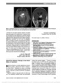
Transient monocular visual loss (TMVL) usually is due to hypoperfusion of the optic nerve or retinal circulation. After the exclusion of thromboembolic and carotid artery diseases, retinal vasospasm should be considered as an underlying cause of TMVL. We report a patient with an increasing number of transient attacks of unilateral blind-ness. Vasospasm was confirmed as the etiology by fundus photography during an attack. Nifedipine 10 mg/d decreased the severity of the visual loss and the number of attacks. The patient was relieved of symptoms entirely with a nifedipine dose of 20 mg/d. Journal of Neuro-Ophthalmology 2014;34:386-388 doi: 10.1097/WNO.0000000000000144 © 2014 by North American Neuro-Ophthalmology Society Transient monocular visual loss (TMVL) often is due to hypoperfusion of the optic nerve or retinal cir-culation (1). It may be caused by embolic disease or atherosclerosis of the carotid arteries (2,3). Treatment typically includes anticoagulation or antiplatelet therapy. In patients with significant internal carotid artery stenosis, carotid artery endarterectomy may be considered (3). After exclusion of thromboembolic and carotid artery diseases, retinal vasospasm should be considered as a poten-tial cause of TMVL. There are several reports of successful treatment of vasospastic TMVL with calcium channel blockers (4-6). CASE REPORT A 28-year-old healthy woman presented to emergency care with transient loss of vision in her right eye. She had experienced complete visual loss in the right eye lasting approximately 2 minutes. The central visual field had recovered first, followed by the gradual recovery of the entire visual field. For the previous 2 days, she had experienced transient irregular scotomas in her right visual field and some pain during eye movement. She had accidentally scratched her right eye with her finger before the onset of symptoms. The patient took no regular medications. She had given birth to her third child 4 months earlier complet-ing a normal pregnancy and delivery. During the past 4 months, had she slept less than normally, only 2 to 6 hours per night, due to childcare. During the same period of time, she experienced severe right-sided head-aches. She had a strong family history of migraine, and one of her siblings reported migraine headaches with visual aura. The patient was admitted to hospital. Visual acuity was 20/20, right eye and 20/25, left eye. The pupils were symmetric and equally responsive to light, and ocular motility was normal. Slit-lamp examination showed a healed epithelial erosion of the right cornea. Dilated funduscopy was normal (Fig. 1). No retinal emboli were observed, and there was no asymmetry between color of the left and right fundus. Results of the neurological examination were normal. Fluorescein angiography of the right fundus was normal. Magnetic resonance imaging of the brain and orbits and the magnetic imaging angiography were unremarkable. Labo-ratory testing showed no signs of inflammatory disease or hypercoagulability and complete blood count, and blood glucose were within normal limits. The patient was pre-scribed acetylsalicylic acid (100 mg/d). The day after admission, the patient experienced 8 episodes of transient loss of vision in her right eye. Duration of the episodes ranged from a few seconds to 5 minutes, and there was a total loss of vision during each attack. The patient was being examined during 1 episode, and a right relative afferent pupillary defect was detected. After the episode, pupillary reactions normalized and kinetic Department of Ophthalmology, Oulu University Hospital, Oulu, Finland. The authors report no conflicts of interest. Address correspondence to Ville Saarela, MD, PhD, Department of Ophthalmology, Oulu University Hospital, Box 21, Oulu 90029, Finland; E-mail: ville.saarela@oulu.fi 386 Pitkänen and Saarela: J Neuro-Ophthalmol 2014; 34: 386-388 Clinical Observation Copyright © North American Neuro-Ophthalmology Society. Unauthorized reproduction of this article is prohibited. perimetry and Hardy-Rand-Rittler and Farnsworth Panel D-15 color perception tests were normal in both eyes. On the following day, the patient had 8 more episodes of TMVL in the right eye. During an attack, the central retinal artery constricted confirming vasospasm (Fig. 2). After the attack, the retinal vessels seemed dilated (Fig. 3). The patient was asymptomatic between the episodes. Nifedipine was prescribed at 10 mg/d. A low dose of nifedipine was chosen because of the patient's relatively low blood pressure (100/65 mm Hg). After the first dose, the patient had only 1 more transient attack of total blindness of the right eye. During the next week, she experienced only a few episodes of partial loss of vision in the right eye, described as dimming or darkening of the periferal visual field. However, the episodes increased in number after the first week (Fig. 4), and the dose of nifedipine was increased to 20 mg/d. With this dose, the patient had no more epi-sodes of TMVL. Her blood pressure remained stable (102/ 60 mm Hg). Seven months after her first attack, the patient became pregnant, and nifedipine was safely discontinued. She has remained symptom-free for over 3 years of follow-up. DISCUSSION Vasospasm has been defined as an inappropriate constric-tion or insufficient dilation in microcirculation (7). Retinal vasospasm should be considered as a possible etiology of TMVL, particularly in young patients with a history of migraine and Raynaud phenomenon. Emotional stress, cold, and physical exercise have been considered to be trig-gering factors of vasospasm (7), and it has been reported to occur more frequently in women of child-baring age (8). Vasospasm may sometimes be demonstrated by angiog-raphy, and vessel changes in the conjunctiva may be present (7). There are photographically documented reports of ret-inal vascular changes during an episode of vasospastic TMVL (4,5,9-17). Calcium channel blockers seem efficacious in treating TMVL due to vasospasm (4). They improve blood flow by reducing the vasoconstrictive effect of endothelin-1 (18). However, the effect of calcium channel blockers is based on case reports and case series (4,5,13,14,19-21). In a num-ber of these reports, different calcium channel blockers have been used at different daily doses. Verapamil has been re-ported effective at doses of 80-120 mg (4,5), nifedipine at FIG. 1. Before episodes of transient visual loss in the right eye, the fundi are normal. FIG. 2. Vasoconstriction of the retinal arteries during an episode of transient monocular visual loss in the right eye. FIG. 3. The retinal arteries and veins appear dilated imme-diately after the episode. Pitkänen and Saarela: J Neuro-Ophthalmol 2014; 34: 386-388 387 Clinical Observation Copyright © North American Neuro-Ophthalmology Society. Unauthorized reproduction of this article is prohibited. doses of 30-80 mg (4,13), and cyclandelate at a dose of 1200 mg (14). The therapeutic effect may be dose-dependent as in our patient. Patients with vasospasm often have low blood pressure, especially during the night (7), and blood pressure should be monitored when starting treatment. The visual prognosis after vasospastic TMVL seems to be good in most patients (22). However, cases of retinal infarc-tion have been reported even in young adults (23). REFERENCES 1. Caplan LR, Hertzer NR. The management of transient monocular visual loss. J Neuroophthalmol. 2005;25:304-312. 2. Petzold A, Islam N, Hu HH, Plant GT. Embolic and nonembolic transient monocular visual field loss: a clinicopathologic review. Surv Ophthalmol. 2013;58:42-62. 3. Benavente O, Eliasziw M, Streifler JY, Fox AJ, Barnett HJ, Meldrum H; North American Symptomatic Carotid Endarterectomy Trial Collaborators. Prognosis after transient monocular blindness associated with carotidartery stenosis. N Engl J Med. 2001;345:1084-1090. 4. Winterkorn JM, Kupersmith MJ, Wirtschafter JD, Forman S. Brief report: treatment of vasospastic amaurosis fugax with calcium-channel blockers. N Engl J Med. 1993;329: 396-398. 5. Bernard GA, Bennett JL. Vasospastic amaurosis fugax. Arch Ophthalmol. 1999;117:1568-1569. 6. Inoue T, Shimizu H, Okabe H, Tominga T. Amaurosis fugax caused by retinal artery vasospasm. Case illustration. J Neurosurg. 2007;106:934. 7. Flammer J, Pache M, Resink T. Vasospasm, its role in the pathogenesis of diseases with particular reference to the eye. Prog Retin Eye Res. 2001;20:319-349. 8. Flammer J, Prünte C. Ocular vasospasm. Functional circulatory disorders in the visual system, a working hypothesis [Article in German]. Klin Monbl Augenheilkd. 1991;198:411-412. 9. Wolter J, Burchfield W. Ocular migraine in a young man resulting in unilateral blindness and retinal edema. J Pediatr Ophthalmol. 1971;8:173-176. 10. Humphrey W. Central retinal artery spasm. Ann Ophthalmol. 1979;11:877-881. 11. Kline L, Kelly C. Ocular migraine in a patient with cluster headaches. Headache. 1980;20:253-257. 12. Burger S, Saul R, Selhorst J, Thurston S. Transient monocular blindness caused by vasospasm. N Engl J Med. 1991;325: 870-873. 13. Petzold A, Islam N, Plant GT. Videoreconstruction of vasospastic transient monocular blindness. N Engl J Med. 2003;348:1609-1610. 14. Heckmann J, Gaul C, Neundorfer B, Harazny J, Michelson G. Vasospastic amaurosis fugax. J Neurol Neurosurg Psychiatry. 2003;74:149. 15. Killer HE, Forrer V, Flammer J. Retinal vasospasm during an attack of migraine. Retina. 2003;23:253-254. 16. Doyle E, Vote BJ, Casswell AG. Retinal migraine: caught in the act. Br J Ophthalmol. 2004;88:301-302. 17. Stasi K, Ramchandran V, Rao NA, Feldon SE, DiLoreto DA Jr. Retinal arteriolar spasm during transient monocular visual loss in eosinophilic vasculitis. J Neuroophthalmol. 2009;29:58-61. 18. Meyer P, Lang MG, Flammer J, Lüscher TF. Effects of calcium channel blockers on the response to endothelin-1, bradykinin and sodium nitroprusside in porcine ciliary arteries. Exp Eye Res. 1995;60:505-510. 19. Ploner C, Valdueza J, Villringer A. Vasospastic amaurosis fugax in a patient with overlap collagenosis treated with nimodipine. Neurol Res. 1995;17:66-69. 20. Teman A, Winterkorn J. Transient monocular blindness associated with sexual intercourse. N Eng J Med. 1995;333:393-394. 21. Winterkorn JMS, Teman A. Recurrent attacks of amaurosis fugax treated with calcium channel blocker. Ann Neurol. 1991;30:423-425. 22. Gautier JC. Amaurosis fugax. N Engl J Med. 1993;329:426-428. 23. Tippin J, Corbett JJ, Kerber RE, Schroeder E, Thompson HS. Amaurosis fugax and ocular infarction in adolescents and young adults. Ann Neurol. 1989;26:69-77. FIG. 4. The number of episodes of TMVL reported by the patient. *On the 15th day, the patient did not record the number of attacks. TMVL, transient monocular visual loss. 388 Pitkänen and Saarela: J Neuro-Ophthalmol 2014; 34: 386-388 Clinical Observation Copyright © North American Neuro-Ophthalmology Society. Unauthorized reproduction of this article is prohibited. |