| OCR Text |
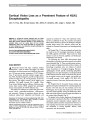
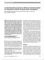
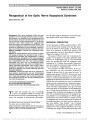
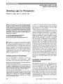
Show Fluorescein Angiographic Identification of Optic Disc Drusen With and Without Optic Disc Edema Stacy L. Pineles, MD, Anthony C. Arnold, MD Background: The fluorescein angiographic criteria for differ-entiating optic disc drusen (ODD) from optic disc edema have been unclear. We designed a study to identify distinguishing angiographic features of each and to apply them to cases where both drusen and edema were present. Methods: A computer search was performed for cases evaluated in a university academic neuro-ophthalmology consultative practice and coded as ODD; all cases were reviewed, and those with fluorescein angiography were selected for further study. Cases were classified as either buried or surface ODD. Ten cases with papilledema were selected for comparison. Eight cases of coexistent drusen and edema were identified. Autofluorescence, early leak-age, early blockage, early and late nodular staining, late peripapillary staining, and late leakage were tabulated. Results: Two hundred sixteen cases of ODD were identified; 62 (116 eyes) had adequate fluorescein angiography for study. Twenty-three eyes were classified as surface ODD; 90% demonstrated early nodular staining of the disc, with late nodular staining in 90% and late circumferential peripapillary staining in 22%; autofluorescence was visible in 93% with preinjection photography. Eighty-three eyes were classified as buried ODD; 25% demonstrated early nodular staining, with late nodular staining in 29% and late circumferential peripa-pillary staining in 80%; autofluorescence was visible in 12% of those with preinjection photography. In 9 eyes, buried ODD were present with superimposed true edema. In these eyes, early dye leakage, late nodular hyperfluorescence, and late leakage were present. Conclusion: Early and late fluorescein angiographic features reliably distinguish ODD from edema and may be particu-larly useful when the conditions coexist. Journal of Neuro-Ophthalmology 2012;32:17-22 doi: 10.1097/WNO.0b013e31823010b8 © 2012 by North American Neuro-Ophthalmology Society The differentiation of optic disc drusen (ODD) from optic disc edema (ODE) is of critical importance be-cause ODE may be a manifestation of a life-threatening condition requiring urgent patient evaluation, whereas ODD is most often a benign process requiring only obser-vation. In cases of ODD located on the disc surface, diagnosis may be straightforward, but with buried ODD, the optic disc appearance may mimic that of ODE. Fur-thermore, ODE may develop in cases with preexisting ODD, making the correct diagnosis even more uncertain. Ancillary testing has been utilized to aid in the identification of ODD, including B-mode ultrasonography (1), CT, and autofluorescence (2-5) Optical coherence to-mography (OCT) has been recently used to identify changes in retinal nerve fiber layer thickness associated with ODD (6) and to attempt to differentiate ODD from ODE (7). Each of these techniques has significant limitations. They are used primarily to confirm the presence of ODD, which does not rule out the coexistent ODE. Fluorescein angiography (FA) features in ODD have been reported pre-viously (5), but specific criteria for differentiating ODD from ODE remain unclear, and the distinction in cases of superimposed ODE has not been addressed. In practice, late-phase FA may be useful in differentiation but is often misinterpreted, with specifics of intrapapillary vs peripapil-lary abnormalities and of dye leakage vs staining of the tissues frequently confused. The early phases of the angio-gram, which aid in the differentiation, are rarely integrated into the analysis. We reviewed FA findings in cases of ODD evaluated at the Jules Stein Eye Institute, comparing fea-tures to ODE and clarifying those features that allow iden-tification of both when they coexist. METHODS This retrospective medical record review was approved by the University of California, Los Angeles, Institutional Review Board. A computer search was performed on all patients evaluated in the consultative neuro-ophthalmology Jules Stein Eye Institute, UCLA Department of Ophthalmology, Los Angles, California. Supported in part by an unrestricted grant from the Research to Prevent Blindness, Inc. Presented at the Annual Meeting of the North-American Neuro- Ophthalmology Society, 2009, Lake Tahoe, NV. The authors report no conflicts of interest. Address correspondence to Anthony C. Arnold, MD, Jules Stein Eye Institute, 100 Stein Plaza, Los Angles, CA 90095-7002; E-mail: arnolda@ucla.edu Pineles and Arnold: J Neuro-Ophthalmol 2012; 32: 17-22 17 Original Contribution Copyright © North American Neuro-Ophthalmology Society. Unauthorized reproduction of this article is prohibited. practice by 1 investigator (A.C.A.) at the Jules Stein Eye Institute from 1990 to 2009, coded as ODD. All cases were examined, and those with fluorescein angiographic studies, performed by routine technique, were selected for further evaluation. Inclusion/Exclusion Criteria ODD were diagnosed clinically (based either on visible ODD on the disc surface or on the presence of blurred/ scalloped optic disc margin and disc elevation without the presence of optic nerve hyperemia, microvascular abnor-malities, or nerve fiber layer edema) or by B-mode ultrasonography (40 eyes) or orbital CT scanning (11 eyes). ODD that were initially diagnosed solely by angiography were excluded. Method of Record Review Color fundus photographs and FAs were reviewed. FAs were evaluated for the following characteristics: 1) the presence or absence of autofluorescence in those angiograms that included preinjection photographs, 2) the presence or absence of early blockage, 3) the presence and characteristics of early TABLE 1. Fluorescein angiographic characteristics of 116 eyes harboring ODD Surface ODD (%) Buried ODD (%) Combined ODD/ODE (%) Autofluorescence 93 12 0 Early blockage 35 11 0 Early nodular staining 90 25 33 Late nodular staining 90 29 100 Late circumferential peripapillary staining 21 8 100 Early leakage 0 0 75 Late leakage 0 0 100 FIG. 1. Surface ODD. A. Drusen visible superiorly and nasally (arrows). B. Arterial phase of FA revealing early blockage of fluorescence due to the presence of surface drusen (arrows). C. Mid-phase angiography revealing early nodular staining of drusen (arrow). D. Late-phase angiogram revealing further nodular staining of drusen (arrows). 18 Pineles and Arnold: J Neuro-Ophthalmol 2012; 32: 17-22 Original Contribution Copyright © North American Neuro-Ophthalmology Society. Unauthorized reproduction of this article is prohibited. fluorescence, 4) the presence and characteristics of late fluorescence, and 5) the presence of dye leakage from the optic disc at any point during the angiogram. Clinical features were tabulated, and ancillary testing was recorded. The records and FA of 10 diagnosed with papilledema due to idiopathic intracranial hypertension (IIH) were reviewed for comparison. RESULTS Patients Two hundred sixteen patients with the diagnosis of ODD were identified. Of those patients, 70 had undergone FA. Sixty-two patients (116 eyes) met inclusion criteria of the study with age ranging from 9 to 72 years (mean, 36 years). Thirty-five patients were women, and 27 were men. Seven of the patients had been treated for IIH prior to our consultation due to the presumed presence of papilledema. Fluorescein Angiographic Findings Fluorescein angiographic features are summarized in Table 1. Twenty-three eyes (20%) were classified as surface ODD, based on the criteria of visible superficial ODD and no clin-ical evidence of ODE (Fig. 1A). Early blockage was seen in 8 eyes (35%) (Fig. 1B); twenty eyes (90%) demonstrated early nodular staining of the optic disc (Fig. 1C). Late nod-ular staining of the disc occurred in 20 eyes (90%) (Fig. 1D). Five eyes (22%) demonstrated late hyperfluorescent staining of the peripapillary area not associated with peripapillary at-rophy (Fig. 2). Autofluorescence was visible in 13 (93%) of the 14 eyes with preinjection photographs. None of the eyes demonstrated early or late leakage of the dye from the disc. Eighty-three eyes (72%) were classified as buried ODD, based on criteria of no visible superficial ODD, elevation and blurred margins of the disc, and no peripapillary retinal vascular obscuration or other clinical evidence of ODE (Fig. 3A). Twenty-one eyes (25%) demonstrated early nodular staining of the optic disc (Fig. 2B); the remaining eyes did FIG. 2. Example of buried ODD (A) with circumferential peripapillary staining in late-phase angiogram (B). FIG. 3. Buried ODD of different patients. A. Blurring of disc margins without blood vessel obscuration or nerve fiber layer opacification. B. Mid-phase angiogram revealing early nodular staining (arrows) of the optic nerve. C. Late-phase angiogram showing late nodular staining (arrow) of the optic nerve. Pineles and Arnold: J Neuro-Ophthalmol 2012; 32: 17-22 19 Original Contribution Copyright © North American Neuro-Ophthalmology Society. Unauthorized reproduction of this article is prohibited. not demonstrate abnormal early disc staining. Early block-age was seen in 9 eyes (11%). Late nodular staining of the disc occurred in 24 eyes (29%) (Fig. 3C). Sixty-six eyes (80%) demonstrated late circumferential peripapillary stain-ing (Fig. 3D). Autofluorescence was visible in 6 of the 49 eyes (12%) with preinjection photographs. None demon-strated early or late leakage of the dye from the disc. Nine eyes (8%) were classified as buried ODD with evidence of superimposed ODE, based on the presence of visible buried ODD, ultrasonographic, or CT confirmation, in addition to peripapillary retinal vascular obscuration by nerve fiber layer opacity (Fig. 4A). Etiologies of the ODE included diabetic papillopathy (2 eyes), ischemic optic neu-ropathy (3 eyes), and IIH (2 eyes). In 2 eyes, no etiology for ODE was detected. Six eyes demonstrated early leakage, 2 of which also showed superimposed early nodular staining (Fig. 4B); 3 eyes showed early nodular staining without leakage. No eyes showed early blocking. All eyes demon-strated late leakage and nodular staining (Fig. 4C). Prein-jection photography was not performed in any of these cases. An example of a patient with coexistent ODD and ODE is shown in Figure 4. A second example from this category of patients is depicted also in Figure 4. This patient presented with visible optic disk drusen, but it was unclear if optic nerve edema was present as well (Fig. 5A). FA revealed nodular staining with focal leakage in both the early (Fig. 5B) and late (Fig. 5C) phases of the angiogram. The ODE was felt to be due to nonarteritic anterior ische-mic optic neuropathy (NAION). Ten eyes with ODE secondary to IIH were analyzed for comparison. All demonstrated early and late diffuse leakage from the optic disc without visible nodularity or blocking patterns. DISCUSSION When ODD are visible on the optic disc surface, identi-fication is straightforward, although it does not rule out the presence of superimposed ODE. The clinical features of buried ODD include optic disc elevation, blurred optic disc margins without obscuration of peripapillary retinal vessels, and nodular border of the optic disc, all in the absence of features of ODE, including retinal nerve fiber opacification with obscuration of retinal vessels, microvascular FIG. 4. Optic nerve with coexistent ODD and ODE. A. Presence of peripapillary nerve fiber layer opacifications with blood vessel obscuration and ODD, characterized by scalloping of the optic disc border. B. Mid-phase angiogram demonstrating early nodular staining (arrow) of the optic disc with minimal leakage. C. Late-phase angiogram showing fluorescein leakage and nodular staining. D. Follow-up photograph 3 years later revealing the presence of ODD without ODE. 20 Pineles and Arnold: J Neuro-Ophthalmol 2012; 32: 17-22 Original Contribution Copyright © North American Neuro-Ophthalmology Society. Unauthorized reproduction of this article is prohibited. abnormalities, such as optic disc surface capillary net dilation, telangiectasia, retinal hemorrhages, and exudates. At times, it may be difficult to distinguish ODD from ODE with certainty. In our case series, 7 patients were treated for IIH but were later found to have ODD as their sole diagnosis. Of the original 206 ODD patients reviewed, 33 underwent CT scanning, 66 underwent MRI, and 24 underwent lumbar puncture. Given the expense and potential risks of these additional tests, the value of simpler less invasive testing to identify ODD and rule out ODE is essential. The detection of autofluorescence of the optic disc on preinjection photography is confirmatory for ODD, but the technique is most effective when the ODD are on or near the disc surface. For buried ODD, sensitivity is low; Kurz- Levin and Landau (1) documented autofluorescence in only 15 of 82 cases (18%) of buried ODD. CT is limited not only by 1.5-mm thickness of orbital sections, which may miss ODD, but also by the fact that calcification must be present for their detection. B-mode ultrasonography simi-larly detects only calcified ODD. While no study has clearly identified the percentage of ODD that are calcified, Kurz- Levin and Landau (1) found positive ultrasonography in only 39 of 82 of eyes (48%) with buried ODD. For this reason, we did not require a positive result on ultrasonog-raphy or CT for confirmation of the diagnosis. In our study, 40 eyes with buried ODD underwent B-mode ultrasonog-raphy; of which, 24 (60%) were positive for calcified ODD. These data corroborate previous studies confirming the lim-ited role for ultrasonography in detection of buried ODD. Johnson et al (7) have reported on the use of time-domain OCT to differentiate ODD from ODE, based on the inter-nal optic nerve contour and the subretinal hyporeflective space. While this technique shows promise, it seems most effective in cases where surface ODD are clinically apparent; in more subtle cases, the distinguishing OCT features are less clear. Buried ODD can be detected with spectral-domain OCT (8), but differentiating ODD from ODE has yet to be established with this technique. FA has been employed to identify ODD and differen-tiate from ODE, but a comprehensive set of criteria, utilizing the entire angiographic sequence, has not been clearly identified. Sanders and Ffytche (5), and Mustonen and Nieminen (4) reported on FA findings in ODD, describ-ing "early fluorescence" and "nodular well-demarcated late hyperfluorescence" seen without leakage. Cartlidge et al (9) compared the FA findings of eyes diagnosed with "pseudo-papilledema" (the percentage with ODD not given) to those of eyes with papilledema, emphasizing the "increased vas-cularity" seen more often in papilledema. Other FA studies (10-12) have differentiated ODD by noting "disc staining" from ODE characterized by "disc leakage." But a clear dis-tinction between hyperfluorescence, staining, and leakage, a critical appraisal of intrapapillary vs peripapillary hyper-fluorescence, and a comparison of the complete FA sequence in distinguishing between ODD and ODE has not been published. Our findings demonstrate that careful assessment of the entire FA can reliably differentiate ODD from ODE. Surface ODD do not require FA for diagnosis. In cases of suspected coexistent ODE, the appearance of early or late leakage confirms the presence of ODE. Buried ODD are characterized by either no early staining (75%) or a characteristic early nodular staining (25%), unlike ODE, which will demonstrate early diffuse leakage. Buried ODD also often show late peripapillary staining, either nodular (29%) or circumferential (80%) or both, which is not seen in ODE. On FA, ODE lead to early and late fluorescein leakage from the optic disc (13,14). The coexistence of ODD and ODE has been described in anecdotal cases of IIH and NAION (15,16), but FA criteria have not been reported. Our data indicate that the presence of fluorescein leakage indicates ODE, and further patient evaluation may be warranted. FIG. 5. Optic nerve with coexistent ODD and ODE. A. The optic nerve has surface drusen (arrows), but the presence of edema is uncertain. B. Arterial phase angiogram demonstrating early nodular staining of the disc with focal fluorescein leakage (arrow). C. Mid-phase angiogram revealing nodular staining of the disc with more fluorescein leakage. Pineles and Arnold: J Neuro-Ophthalmol 2012; 32: 17-22 21 Original Contribution Copyright © North American Neuro-Ophthalmology Society. Unauthorized reproduction of this article is prohibited. We recognize the limitations of our study including its retrospective nature, which may have led to a bias toward obtaining fluorescein angiograms in atypical cases and in those in whom the diagnosis of ODD was more problem-atic. Nevertheless, our data indicate that in patients with surface or buried ODD suspected of also having ODE, full-sequence FA analysis plays a valuable role in establishing the correct diagnosis. REFERENCES 1. Kurz-Levin MM, Landau K. A comparison of imaging techniques for diagnosing drusen of the optic nerve head. Arch Ophthalmol. 1999;117:1045-1049. 2. Friedman A, Beckerman B, Gold DH, Walsh JB, Gartner S. Drusen of the optic disc. Surv Ophthalmol. 1977;21: 375-390. 3. Kelley J. Autofluorescence of drusen of the optic nerve head. Arch Ophthalmol. 1974;92:263-264. 4. Mustonen E, Nieminen H. Optic disc drusen-a photographic study I. Autofluorescence pictures and fluorescein angiography. Acta Ophthalmol. 1982;60:849-858. 5. Sanders MD, Ffytche TJ. Fluorescein angiography in the diagnosis of drusen of the disc. Trans Ophthalmol Soc U K. 1968;87:457-468. 6. Katz BJ, Pomeranz HD. Visual field defects and retinal nerve fiber layer defects in eyes with buried optic nerve drusen. Am J Ophthalmol. 2006;141:248-253. 7. Johnson L, Diehl ML, Hamm CW, Sommerville DN, Petroski GF. Differentiating optic disc edema from optic nerve head drusen on optical coherence tomography. Arch Ophthalmol. 2009;127:45-49. 8. Yi K, Mujat M, Sun W, Burnes D, Latina MA, Lin DT, Deschler DG, Rubin PA, Park BH, de Boer JF, Chen TC. Imaging of optic nerve head drusen: improvements with spectral domain optical coherence tomography. J Glaucoma. 2009;18:373-378. 9. Cartlidge N, Ng RC, Tilley PJ. Dilemma of the swollen optic disc: a fluorescein retinal angiography study. Br J Ophthalmol. 1977;61:385-389. 10. Arnold AC. Optic disc drusen. Ophthalmol Clin North Am. 1991;4:505-517. 11. Auw-Haedrich C, Staubach F, Witschel H. Optic disk drusen. Surv Ophthalmol. 2002;47:515-532. 12. Brodrick JD. Drusen of the disc and retinal haemorrhages. Br J Ophthalmol. 1973;57:299-306. 13. Miller S, Sanders MD, Ffytche TJ. Fluorescein fundus photography in the detection of early papilledema and its differentiation from pseudo-papilledema. Lancet. 1965;2:651-654. 14. Oosterhuis J, Boen-Tan TN. Fluorescein angiography in papilledema and pseudo-papilledema. Ophthalmologica. 1969;159:96-110. 15. Karel I, Otradovec J, Peleska M. Fluorescence angiography in circulatory disturbances in drusen of the optic disk. Ophthalmologica. 1972;164:449-462. 16. Liew SC, Mitchell P. Anterior ischaemic optic neuropathy in a patient with optic disc drusen. Aust N Z J Ophthalmol. 1999;27:157-160. 22 Pineles and Arnold: J Neuro-Ophthalmol 2012; 32: 17-22 Original Contribution Copyright © North American Neuro-Ophthalmology Society. Unauthorized reproduction of this article is prohibited. [KBDopticnervedrusen] |