| OCR Text |
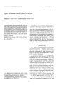
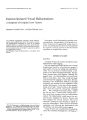
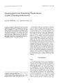
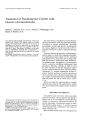
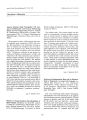
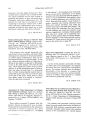
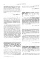
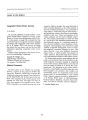
Show Journal of Clinical Neuro-ophthalmology 8(2): 99-104, 1988. Occult Pituitary Apoplexy Associated with Craniopharyngioma Rodney I. Kellen, M.B., B.C.H., MMed, F.R.C.S., Ronald M. Burde, M.D., and Fred J. Hodges, III, M.D. © 1988 Raven Press, Ltd., New York Craniopharyngiomas generally are not considered in the differential diagnosis of pituitary apoplexy. A case of craniopharyngioma is presented, which emphasizes the need for magnetic resonance imaging in the setting of overt or subclinical pituitary apoplexy. Key Words: Craniopharyngiomas-Pituitary apoplexy. From the Departments of Ophthalmology (R.I.K., R.M.B.), Neurology and Neurological Surgery (R.M.B.), and the Malhnkcrodt Institute of Radiology (F.}.H.), Washington UniverSity School of Medicine, St. Louis, Missouri. Address correspondence and reprint requests to Ronald M. Burde, M.D., Department of Ophthalmology, Montefiore MedIcal Center, 111 East 210 Street, Bronx, NY 10467, U.S.A. 99 In nonobstetric cases, pituitary apoplexy implies sudden hemorrhagic necrosis of the pituitary gland associated with pituitary tumor. David and associates (1) regard pituitary apoplexy as a syndrome produced by any sudden enlargement of a preexisting pituitary adenoma. The onset of this event is usually heralded by the classic triad of headache, oculomotor or afferent visual system dysfunction, and evidence of subarachnoid hemorrhage. In the past, this condition was always considered to be an emergency, requiring urgent surgical decompression to prevent a fatal outcome or at least to minimize resultant morbidity (2-4). More recently, lesser forms of pituitary apoplexy have been described that were successfully managed without surgical intervention (5-8). Thus, it is appropriate to consider pituitary apoplexy as a state with a spectrum of severity ranging from subclinical to rapidly fatal (7). We recently saw a patient who presented with an occult hemorrhage in a preexisting craniopharyngioma following minor head trauma. CASE REPORT A 37-year-old white man was diagnosed as having a craniopharyngioma at age 15. He presented at that time with a history of diplopia and was found to have external ophthalmoplegia and hydrocephalus. Radiographic studies demonstrated intracranial parasellar calcification. Despite a lack of tissue diagnosis, the sellar region was irradiated (dose unknown). Until October of 1986, the patient did well with stable visual acuities of 20/15 in both eyes and normal visual fields and extraocular motility. He did, however, exhibit endocrine dysfunction, i.e., he was impotent with low serum testosterone, and he also required mainte- OCCULT PITUITARY APOPLEXY 101 FIG. 1. Midsagittal T,-weighted image showing high (white), intermediate (gray), and low (black) signal intensity making up the intra- and suprasellar mass. The high signal below the mass posteriorly is due to the fatty bone marrow of the clivus, while the absent signal beneath the sella represents air in the sphenoid sinus (T, = TR 500, TE 15). tuitary apoplexy using CT scans and believed they could identify blood within an adenomatous pituitary gland. They were further able to discriminate between acute and chronic hemorrhage. In unenhanced scans they noted soft-tissue masses with acute hemorrhage or areas of low density alone indicating chronic hemorrhage or necrosis. With contrast there was no enhancement, partial enhancement, uniform enhancement, or ring-like enhancement. Post et al. did, however, recognize that some cases of both acute and chronic hemorrhage could be overlooked. Fujimoto and associates (15) described a density level of fluid blood within a ring-enhancing lesion, which they believed was almost certain evidence of pituitary apoplexy within a pituitary adenoma. In the presence of a prominent episode of pituitary apoplexy the mere presence of a large sellar mass was thought to be adequate to confirm the diagnosis (16). In all these cases, angiography was undertaken to exclude a suprasellar aneurysm. The advent of MR imaging has greatly enhanced the physician's ability to detect intracranial pathology, even where CT has failed (17,18). MR imaging is particularly useful in detecting the presence of subacute hemorrhage. Several tissues are known or thought to cause elevated signal in TI-weighted, Tz-weighted, and proton density sequences, including subacute or chronic hemorrhage, cholesteatoma, and mucocele. Mixed signals could, of course, represent a variety of normal and pathologic tissues, but the lucent appearance on CT, in this case, tends to exclude such entities as recent blood clot, intrasellar calcification, and solid pituitary adenoma or meningioma. Cerebrospinal fluid as in empty sella and arachnoid cyst; cystic craniopharyngioma or cystic adenoma; epidermoid, dermoid or teratomatous cyst; inflammatory exudate (abscess); mucocele; and fatty tissue are considerations. Craniopharyngioma may contain cholesterol, keratin nodules, epithelial nests or cords, hemorrhagic products, and necrosis (19). By MR imaging it is frequently cystic appearing (20). High protein J Clill Neuro-ophthaltllol, Vol. 8, No.2, 1988 OCCULT PITUITARY APOPLEXY 103 .. _l> FIG. 3. Photomicrograph of cholesterol clefts (small arrow) with associated degenerating red blood cells (large arrow). Cystic spaces lined by palisading epithelial cells are typical of craniopharyngioma (asterisks) ( x 90). and suprasellar lesion could easily represent hemorrhage 1 or more weeks old. The clinical presentation of our patient with a history of recent minor head trauma associated with decreased vision, abnormal visual fields, and a past history of a para sellar mass suggests sudden expansion of the parasellar mass lesion. Surgical and histopathological confirmation of hemorrhage into the craniopharyngioma is, we feel, supported by the MR findings, despite the lack of absolute precision in defining tissue type by MR imaging. Although craniopharyngioma previously was not thought to be associated with hemorrhagic infarction of the pituitary gland (22), it should be considered in the differential diagnosis of pituitary apoplexy. When acute hemorrhage is clinically suspected in the setting of a known parasellar tumor, CT is more diagnostic in the first week. If the CT is negative or ambiguous, or if the patient presents after 1 week, then MR imaging is the appropriate imaging modality. Acknowledgment: Supported in part by an unrestricted grant from Research to Prevent Blindness, Inc., New York, New York (Department of Ophthalmology). REFERENCES 1. David NJ, Gargano FP, Glaser JS. Pituitary apoplexy in clinical perspective. In: Glaser ]S, Smith ]L, eds: Neuroophthalmology (vol 8). St. Louis: Mosby, 1975:140-65. 2. lrsigler F]. Surgical emergencies related to the pituitary and neighbouring areas. S Afr Med 11960;34:10-4. 3. Zervas NT, Mendelson G. Treatment of acute haemorrhage of pituitary tumours. Lallcet 1975;1:604-5. 4. Robit RL, Fein ]M. Pituitary apoplexy: a review and reappraisal. 1 Nel/rosl/rg 1972;37:280-7. 5. Reid RL, Quigley ME, Yen SSe. Pituitary apoplexy. A review. Arch Nel/roI1985;42:712-9. 6. Tsitsopoulos P, Andrew L Harrison MJG. Pituitary apoplexy and haemorrhage into adenomas. Postgrad Med 1 1986;62:623-6. 7. Peter SA. Subclinical pituitary apoplexy. NY State] Med 1986;86(12):656-7. 8. Wright RL, Ojemann RG, Drew JH. Hemorrhage into pituitary adenomata. Report of two cases with spontaneous recovery. Arch Neurol 1965;12:326-31. 9. Petito CK. DeGirolami U, Earle KM. Craniopharyngiomas. A clinical and pathological review. Callcer 1976;37:1944-52. 10. Kennedy HB, Smith RJS. Eye signs in craniopharyngioma. Br 1 OphthalmoI1975;59:689-95. 11. Isayama Y. Changes in visual field associated with changes of tension in large cyst, which is craniopharyngioma, extending to the middle cranial fossa. Nippoll Ganka Gakkai Zasshi 1970;74(7):596-604. 12. Holness RO, Ogundimu FA, Langille RA. Pituitary apo- 1Cli" NClIro-ophthalmol, Vol. 8, No.2, 1988 104 R. I. KELLEN ET AL. plexy following closed head trauma. Case report. 1Neurosurg 1983;59:677-9. 13. Lloyd MH, Belchetz PE. The clinical features and management of pituitary apoplexy. Postgrad Med 11977;53:82-5. 14. Post MJD, David NJ, Glaser JS, Safran A. Pituitary apoplexy: diagnosis by computed tomography. Radiologl/ 1980;134:665-70. . 15. Fujimoto M, Yoshino E, Ueguchi T, Mizukawa N, Hirakawa. K. Fluid blood density level demonstrated by computenzed tomography in pituitary apoplexy. Report of two cases. JNeurosurg 1981;55:143-4. 16. Haigh SE, Lau DCW, Rorstad OP. Usefulness of computerIzed tomography and cerebral angiography in the diagnosis of pituitary apoplexy [Abstract). Clill l/luest Med 1986;9(suppl):A37. 17. Johnson LN, Hepler RS, Yee RD, Frazee JG, Simons KB. Magnetic resonance imaging of craniopharyngioma. Am J Ophthalmol 1986;102:242-4. 18. Lee BCP, Deck MDF. Sellar and juxtasellar lesion detection with MR. Radiology 1985;157:143-7. 19. Buerger PC Vogel FS. Surgical pathology of the nervous sl/stem and its couerillgs, 2nd ed. New York: Wiley & Sons, 1982:511-25. 20. Karnaze MG, Sartor K, Winthrop JD, Gado MH, Hodges FJ. Suprasellar lesions: evaluation with MR imaging. Rndi% gy 1986;161:77-82. 21. Pusey E, Kortman KE, Flannigan BD, Tsuruda J, Bradley WG. MR of craniopharyngiomas: tumor delineation and characterization. Am 1 Neuroradiol 1987;8:439-44. 22. Case Records of the Massachusetts General Hospital. Case 3-1986. N Ellgll Med 1986;314:229-38. |