| OCR Text |
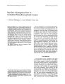
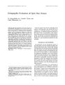
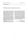
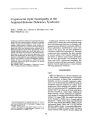
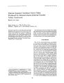
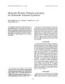
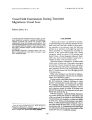
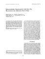
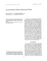
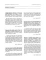
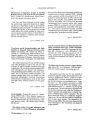
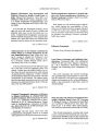
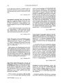
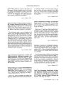
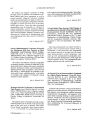
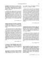
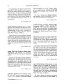
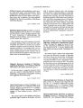
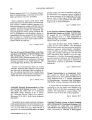
Show Journal of Clinical Neuro- ophthalmology 11( 2): 111- 113, 1991, Monocular Elevation Weakness and Ptosis: An Oculomotor Fascicular Syndrome? Emmanuel Hriso, M. D., Joseph c. Masdeu, M. D., and Aaron Miller, M. D. © 1991 Raven Press, Ltd" New York The topographic arrangement of the fascicular portion of the oculomotor nerve in the midbrain is not known. A patient with infarction involving the lateral portion of the fascicle had isolated monocular elevation paresis and ptosis, suggesting that the fibers destined to the elevators of the eye and eyelid course laterally in the fascicle. Key Words: Eye movements- Oculomotor nerveMesencephalon- Ophthalmoplegia- X- ray computed tomography. From the Departments of Neurology, St. Vincent's Medical Center ( E. H., J. eM.), New York Medical College and New York University School of Medicine a. eM.), Maimonides Medical Center and the State University of New York Health Science Center at Brooklyn ( AM.), and the Albert Einstein College of Medicine a. eM., AM.), New York, New York, U. s. A Address correspondence and reprint requests to Dr. J. e Masdeu, Department of Neurology, St. Vincent's Medical Center, 153 West 11th Street, New York, NY 10011, U. S. A 111 Although the nuclear innervation of the oculomotor muscles has been extensively studied, the topographic representation of the fibers in the fascicular portion of the oculomotor nerve is poorly understood ( 1). We report a patient with a lateral midbrain infarct who had dysfunction restricted to unilateral elevation weakness and ptosis, in the absence of other extraocular muscle abnormalities. This case suggests a lateral location of fascicular oculomotor fibers destined for the levators of the eye and eyelid. CASE REPORT A 75- year- old man with a history of cardiac arrhythmias had a sudden onset of right- sided weakness and dysarthria. On examination, the patient was fully oriented. He had an incomplete left ptosis, and supraduction of the left eye was limited to 15° both on abduction and adduction. Upward excursion was not increased by the oculovestibular reflex. Bell's phenomenon was not tested for. All other eye movements were intact ( Fig. 1). Both pupils were 4 mm in diameter and reacted well to light. There was no nystagmus. His right side was hemiplegic. The remainder of the neurological examination was normal. Computed tomography ( CT) scan on admission revealed a hypodense lesion involving the left cerebral peduncle and the anterior portion of the midbrain tegmentum, sparing the midline structures ( Fig. 2). II- weighted magnetic resonance imaging ( T2 images were technically poor) performed 8 weeks later failed to show any abnormalities. Ocular motility recovered fully in a 6- month period. DISCUSSION In the present case, both clinical and CT findings suggest involvement of the fascicular oculomotor fibers as they transverse the midbrain tegmentum. 112 E. HRISO ET AL. FIG. 1. From top to bottom, primary position, gaze up and to the left. up and to the right. to the right, and down. A supranuclear location is unlikely in the presence of ptosis, hypotropia in the primary position, and a lack of reflex movements ( 2- 4). The unilaterality of the ptosis and elevation weakness militates against nuclear involvement. Nadeau and Trobe ( 5) postulated partial involvement of the fascicular portion of the oculomotor nerve in a patient with similar clinical and CT findings to the one reported here. Also similar were the clinical findings in two of the patients reported by Ksiazek and co- workers ( 6). Unlike their third 1Clin Neuro- ophtlu> lmol, Vol. 11, No 2, 1991 FIG. 2. CT scan obtained 6 hours after the stroke, showing a hypodense lesion ( arrow) involving the left cerebral peduncle and the anterior portion of the midbrain tegmentum. sparing the midline structures. patient, who had a medially located lesion and sparing of the eyelid and eye elevation, a lateral midbrain infarct was documented in a patient with selective ptosis and elevation weakness. Starr ( 7) mentioned a case with autopsy reported by Kahler and Pick in which the levator palpabrae superioris and elevators of the left eye were predominantly affected. A hemorrhage involved the red nucleus and the lateral fascicular oculomotor fibers. These cases suggest that lesions located laterally in the midbrain tegmentum may cause selective involvement of the elevator of the eye and eyelid. Our patient's CT localized the lesion to the lateral portion of the midbrain, spanning the crus cerebri and midbrain tegmentum ( Fig. 2). This area is traversed by the most lateral bundles of the fascicular portion of the oculomotor nerve ( Fig. 3). Axons from the different oculomotor subnuclei leave the anterior aspect of the nucleus, spreading in a widely divergent pattern as they course anteriorly through the red nucleus, and converge near their exit in the interpeduncular fossa. The span of the divergent oculomotor fibers ( approximately 3 mm in the sagittal plane and 6 mm in the axial or transverse plane) is enough to explain selective involvement of the lateral or medial bundles by midbrain pathology. Little data is available on the topographic distribution of the fibers in the fascicle. Fibers destined to the superior rectus originate in the contralateral nucleus of III and cross at the level of the nucleus ( 8,9). In a rendition by Ramon y Cajal of the origin of the fascicular portion of ill in the hawk, the crossing fibers tend to be distributed in the lateral bundles of the fascicle ( Fig. 4) MONOCULAR ELEVAnON PALSY FIG. 3. Transverse midbrain section at the level of the oculomotor nucleus. Crosshatched: area of infarction. Left side: Woelcke's myelin stain of a normal midbrain. Note the wide sweep made by the fascicular fibers as they traverse the red nucleus. The lateral fibers ride on the lateral aspect of the red nucleus ( arrowheads). Right side: diagram of the fascicular portion of the oculomotor nerve. 113 , ' ,. a. 1/' ( _, I FIG. 4. Cajal's rendition of the distribution of the crossing fibers of III ( 0), which course in the lateral bundles of the fascicle ( H). Fledging hawk, reduced silver nitrate method. Reproduced with permission from S. Ramon y Cajal ( 10). ( 10). In humans, a lateral location in the oculomotor fascicle of the fibers destined to the elevators of the eye and eyelid is suggested by the findings in the patient described here. REFERENCES 1. Miyazaki S. Location of motoneurons in the oculomotor nucleus and the course of their axons in the oculomotor nerve. Brain Res 1985; 348: 57- Q3. 2. Barsoum- Homsy M. Congenital double elevator palsy. JPed Ophthalmol Strabismus 1983; 20: 185- 91. 3. Ford CS, Schwartze GM, Weaver RG, Troost BT. Monocular elevation paresis caused by an ipsilateral lesion. Neurology 1984; 34: 1264- 7. 4. Lessell S. Supranuclear paralysis of monocular elevation. Neurology 1975; 25: 1134- Q. 5. Nadeau SE, Trobe JD. Pupil sparing in oculomotor palsy: a brief review. Ann NeuroI1983; 13: 143-- 8. 6. Ksiazek SM, Repka MX, Maguire A, et al. Divisional oculomotor nerve paresis caused by intrinsic brainstem disease. Ann Neurol 1989; 26: 714- 8. 7. Starr MA. Ophthalmoplegia externa partialis. J Nen> Ment Dis 1888; 13: 301- 16. 8. Warwick R. Representation of the extraocular muscles in the oculomotor nuclei of the monkey. J Comp Neurol 1953; 98: 449- 503. 9. Bienfang DC. Crossing axons in the third nerve nucleus. Invest OphthalmoI1975; 14: 927- 31. 10. Ramon y Cajal S. Histologie du systeme nerveux de /' homme et des veriebres. Madrid: Consejo Superior de Investigaciones Cientificas, 1955: 243. JClin Neuro- ophthalmol, Vol. 11, No. 2, 1991 |