| OCR Text |
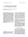
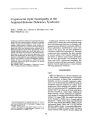
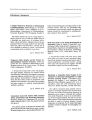
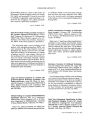
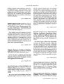
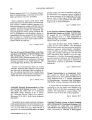
Show ( J 1991 Raven Press, Ltd., New ' 1' 0'" _' ues Pupillary Sparing Oculomotor Nerve Palsy Really Spare the Pupil? Sergiu C. Blumen, M. D., Vera Feiler- Ofry, Amos D. Korczyn, M. D., M. Sc. We examined the pupillary cycle time ( PCT) in eight elderly patients with isolated oculomotor nerve palsy ( ONP) that was characterized by complete involvement of the extraocular muscles. In addition to advanced age, all patients had at least one other vasculopathic risk. fac · tor. Although in all cases the pupil was completely spared by clinical impression, the PCT was significantly prolonged compared with the other eye and well outside the normal range ( mean 1590 .:!: 212 mscc on the involved side and 1076 .:!: 110 on the uninvolved side). On re- examination, after an interval of 2- 3 months. the PeT was either normal or markedly improved in all patients, paralleling the recovery of extraocular muscle function. These findings suggest that even in pure, noncompressive ONPs there is subclinical pupillary involvement. Re~ ated peT examinations may provide an objective means to estimate recovery. Moreover, in the problematic subgroup of " pupil sparing" incomplete ONP, PeT monitoring during the first days may indicate possible progression of a compressive lesion. Key Words: Oculomotor nerve palsy- Pupil- Pupillary cycle time- Parasympathetic nervous system- Autonomic innervation. From the Department of Neurology ( s. c. a., A. D. K.) and Ophthalmology ( V. F- O.), Tel- Aviv Sourasky Medical Center, Sackler Faculty of Medicine, Tel- Aviv University, Israel. Address correspondence and rt'prinl re'lut'sts to Prof. AD Korczyn at Sadder Faculty " I' M~,(! icin('. Tel · Aviv University, Ramal Aviv [, 9978. I~ r" el, M. D., and The assessment of pupillary involvement is crucial in the evaluation of patients presenting with oculomotor nerve palsy ( ONP). Patients with nerve compression, particularly due to posterior communicating artery aneurysms, are likely to have nonreacting--- or at least sluggish- pupils ( 13). On the other hand, when the GNP is due to ischemia, the pupils are rarely involved ( 4,5). The reason for the pupillary sparing is unknown, but it is speculated to be due to the more peripheral 10cation of the parasympathetic fibers in the oculomotor nerve ( 6). On the rare occasions in which compressive lesions produce pupil- sparing ONP, these are usually incomplete ( 3,7,8). As opposed to noncompressive lesions, these may progress over several days to produce the complete paralysis with pupillary involvement ( 7). Unfortunately, the bedside evaluation of pupillary reaction to light depends on clinical impressions and is qualitative rather than quantitative. In an attempt to evaluate whether a more sensitive, quantitative test will disclose some pupillary involvement in patients with ischemic ONP, we have utilized the edge- light pupil cycle time ( PCl) ( 9) in the evaluation of ONP. SUBJECTS AND METHODS Eight patients ( 6 men) with ONP were studied. They had acute onset of complete paralysis of all the muscles innervated by the oculomotor nerve, but an apparently normal pupillary reaction to light and accommodation. The clinical details, shown in Table 1, demonstrate that patients were of advanced age and had other vasculopathic risk factors. The remainder of the neurological and ophthalmological examination was also noncoo. tributory, although patients with diabetes mellitus 92 PCT IN OCULOMOTOR NERVE PALSY 93 TABLE 1. Pupillary cycle time ( peT) in eight patients with " pupil sparing" oculomotor nerve palsy 1st Examination 2nd Examination Abnormal Normal Abnormal Normal Age/ sex Risk factors side side side side E. O. M. 69/ M OM 1533 1000 1200 1000 improved 60/ F OM, HT, lHO 1733 1133 1133 1133 recovered 73/ F HT 1666 1266 1300 1266 recovered 74/ M OM 1266 966 966 966 recovered 74/ M OM, IHO 1933 1133 1200 1133 recovered 781M OM, HT 1533 966 1000 966 recovered 811M OM 1966 1066 1066 1066 recovered 771M OM 1733 1066 1066 1066 recovered Eight pts, mean: 1670 : t 212 1074.:!: 110 1116.:!: 120 1074: tll0 Values are given in milliseconds, mean : t SO. E. O. M" extraocular movements; OM, diabetes mellitus: HT, arterial hypertension: IHO, ischemic heart disease. had impaired vibration sense and no Achilles tendon reflexes. In each case, the peT was measured bilaterally within 48 hours after the onset of GNP and was repeated 2- 3 months later. Pupillary cycle time was measured using a Haag- Streit slit lamp ( Berne, Switzerland), with a horizontal beam of light ( thickness, 0.5 mm) directed at the lower margin of the pupil ( 9,10). Three cycle rounds lasting 30 sec each were produced in each eye, and the average peT was calculated by dividing the 90- sec time period by the total number of cycles elicited. Statistical evaluation was performed using Student's t test for paired samples, comparing the two eyes on the first examination, or the abnormal eye on the two tests. RESULTS When first examined, all patients had complete GNP with apparent nomnvolvement of the pupil. However, the peT was prolonged on the ophthalmoplegic side in each case ( Table l). The difference between the paretic and the uninvolved eye ranged from 300 to 900 msec and was statistically highly significant ( p < 0.01). At the time of the second exarrunation, all patients showed Significant or complete improvement of their extraocular movements. The peT showed recovery or improvement ( Fig. 1). DISCUSSION The peT is an established method for the rapid, quantitative, accurate evaluation of function of the optic and pupillomotor pathways ( 9- 11). The slightly prolonged latencies on the clinically uninvolved side in our patients is consistent with their advanced age ( 11,12). In addition, 7 out of 8 subjects had underlying diabetes mellitus, which also prolongs the peT. The clinical presentation and eventual recovery suggest that in all of our patients ONP was due to ischemia; their age and vasculopathic risk factors are consistent with this diagnosis. All patients underwent brain computerized tomography which did not reveal any structural abnormality that could explain the patients' ophthalmoplegia. Our findings dearly demonstrate that in all eight cases there was subclinical involvement of the parasympathetic component of the oculomotor nerve. Exceptions to the " rule of the pupil" ( 7) are well known; as many as 25% of patients with ischerruc GNP may have clinical evidence of pupil involvement. The present results suggest that subclinical involvement is even more common. In some cases, in whom the rule of the pupil is not obeyed ( 3,7,8), repeated measurements of the peT may be helpful in establishing the diagnosis. For example, in an incomplete ONP, the progressive prolongation of the PCT may indicate an underlying compressive lesion and thus suggest the need for performing angiography. In others, grad- 2000 1800 u ell 1600 < II e - 1400 ~ Il.:. l. I 1200 1000 800 +- 7.":-==~~;;-::-~=-:-;--;:-::-------:---- Normal Rbnormfll Rbnormfll First Second FIG. 1. Pupillary cycle time in patients with " pupil sparing" third nerve palsy. , ( lin Neuro- ephlhalmol, Vol. 11, No, 2, 1991 94 S. C. BLUMEN ET AL. ual improvement of PCT may herald recovery of the ONP, and thus suggest a benign etiology. REFERENCES l. Rucker CW. Par", lysis of the third, fourth and sixth cranial nerves. Arch aphtha/ mol 1958; 46: 787- 94. 2. Green WR, Hackett ER, 5<: hle. tinger N5. NelITa- ophthalmologic evaluation of oculomotor parillysis. Arch Ophtha/ · rna/ 1964; 72: 1~ 7. 3. Kissel JT. Burde RM. Klingele TG. el al. Pupil- sparing oculomotor palsies with internal carotid- posterior communicating artery aneurysms. All" NtwnN 1983; 13: 149- 54. 4. Goldstein JE, Cogan DC. Diabetic ophth. llmoplegia with spedal reference to the pupil. ATd, Oph/ Ira/ mol 1960; 64: 592600 S. Asbury AI<, Aldredge H, Hershberg R, el a!. Oculomotor palsy in diabetes mellitus: A clinicopathologic study. Brain 1970; 83: 555- 66. 6. Kerr FWL, Hollowell OW. Location of pupillomotor and accomodation fibers in the oculomotor nerve. Experimental observations on paralytic mydriasis. JNrurDl NtuTOSllrg Psy · chiatry 1964; 267: 473- 81. 7. Tmbe JD. Third nerve palsy and Ihe pupil. Arch OphlhallTUlJ 1988; 106: 601- 2. 8. Lustbader JM, Miller NR. Painless, pupil- sparing but otherwise complete oculomotor nerve paresis caused by basilar artery aneurysm. Arch Ophthalmoll988; I06: 583. 9. Miller SO, Thompson HS. Edge- light pupil cycle time. Br J Ophthalmoll978; 62: 495- SOO. 10. Martin CN. Edge- light pupil cycle time: a quantifiable autonomic renex IAbstractJ. J Nrllcol Nt: llr05llrg PsychUitry 1985; 48: 6021. 11. Campbell fW, Whiteside TCO. Induced pupillary oscillations. Br JOphthalmoI1950; 34: 180- 9. 12. Manor RS, Yassur Y, Siegal R, Ben- Sira I. The pupil cyde time lesl: age variations in normal patients. Br JOphthalmol 1981; 65: 750-- 3. |