OCR Text |
Show Subretinal Abscess Causing Restricted Diffusion on Magnetic Resonance Imaging Crandall Peeler, MD, Hemant Parmar, MD, Jonathan D. Trobe, MD Background: 
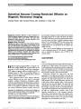
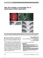
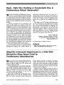
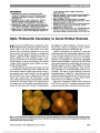
Restricted diffusion of water molecules on diffusion-weighted magnetic resonance imaging most com-monly associated with acute stroke, has also been described in brain abscess. It has been reported in only one case of sub-retinal abscess. Methods: Review of two cases. Results: Two cases of visual loss from subretinal abscess had restricted diffusion in the region of the abscess. In the first case, DWI revealed restricted diffusion in a white mass visible in the posterior retina. In the second case, restricted diffusion was present in an anterior retinal mass invisible by ophthalmoscopy and ultrasound. In combination of restricted diffusion in the cerebrum consistent with septic emboli, these imaging abnormalities allowed earlier treatment that either preserved vision or prevented enucleation. Journal of Neuro-Ophthalmology 2013;33:354-358 doi: 10.1097/WNO.0b013e318294314c © 2013 by North American Neuro-Ophthalmology Society Diffusion-weighted imaging (DWI) is a magnetic reso-nance imaging (MRI) pulse sequence based on the movement of water molecules within brain parenchyma. DWI assigns an apparent diffusion coefficient (ADC) to biological tissues (1). When the extracellular diffusion of water via Brownian motion is impeded by edema, restricted diffusion is seen on DWI (2-8). Most commonly found in acute ischemic infarction, restricted diffusion also has been noted in intracranial neoplastic conditions (lymphoma, meningioma, epider-moid) (1,2) and inflammatory disorders (abscess, empyema, acute demyelinating plaques) (3). In head and neck cancer, DWI has been applied to differentiate benign from malig-nant tumors, discriminate between recurrent tumors and post-therapeutic changes, and detect lymph node metastasis for staging purposes (4). In spine and spinal cord disease, DWI aids in distinction of acute demyelination from infarc-tion and trauma, and helps to identify extradural osseous lesions (5). In orbital disease, DWI has been used to dem-onstrate ischemic and infiltrating optic neuropathies (6), endophthalmitis (7), and thrombosis of the superior oph-thalmic vein (8). Only 1 case of restricted diffusion has been reported in a case of subretinal abscess (9). We describe 2 additional cases to emphasize the diagnostic value of DWI in this unusual setting. CASE REPORTS Case 1 A 16-year-old boy presented with pain and blurred vision in the right eye of 1-day's duration. For the previous 10 days, he had experienced fever, generalized weakness, cough, joint pain, and headaches that were attributed to mononucleosis based on a positive Monospot test. He had received nebu-lizer treatment, ketorolac, and intravenous fluids. Examination disclosed that visual acuity was hand movements in the right eye and 20/20 in the left eye. There were no ocular adnexal abnormalities or tenderness to palpation. The right pupil measured 4 mm and the left pupil measured 2 mm in dim illumination, and there was a right relative afferent pupillary defect. Ocular ductions were full and the eyes were aligned. The visual field was unobtainable in the right eye due to poor visual acuity and full to finger counting in the left eye. Slit-lamp biomicro-scopy and intraocular pressures were normal in both eyes. In the right eye, ophthalmoscopy revealed a large (3 disc diameters horizontally by 4 disc diameters vertically), yellow-white, round subretinal lesion with indistinct mar-gins. In the left eye, there was a smaller (1/2 disc diameter) subretinal fluid collection nasal to the optic disc. Fundus photography was not performed at that time. Departments of Ophthalmology and Visual Science, Kellogg Eye Center (CP, JDT), Radiology, Division of Neuroradiology (HP), and Neurology (JDT), University of Michigan, Ann Arbor, MI. The authors report no conflicts of interest. Address correspondence to Jonathan D. Trobe, MD, Department of Ophthalmology and Visual Science, Kellogg Eye Center, 1000 Wall Street, Ann Arbor, MI 48105; E-mail: jdtrobe@med.umich.edu 354 Peeler et al: J Neuro-Ophthalmol 2013; 33: 354-358 Original Contribution Copyright © North American Neuro-Ophthalmology Society. Unauthorized reproduction of this article is prohibited. MRI of the brain and orbits was performed on a 1.5 T magnet (Siemens Medical Systems, Enlargen, Germany) using an 8-channel head coil. Diffusion imaging data were obtained using an echo-planar single-shot technique with the shortest TR, 89 ms TE, and a 90 flip angle, and with a b value of 1,000 seconds/mm2. The data were recorded on a 128·128 matrix and were zero filled for a final resolution of 128·256. Axial slices with 5-mm slice thickness and a 0.5-mm interslice gap were obtained. DWI revealed a small nodular abnormality in the posterior aspect of the right globe that exhibited restricted diffusion (Fig. 1). In addition, multiple foci of restricted diffusion were visualized in both cerebral and cerebellar hemispheres in cortical, subcortical, and periventricular lo-cations (Fig. 2A). Most of these foci exhibited dark signal on the corresponding ADC maps, confirming that the DWI signal reflected true areas of restricted diffusion without any T2 effect (Fig. 2B). Susceptibility sequences demonstrated punctate hemorrhagic components within some of the brain lesions. After contrast administration, many of the lesions demonstrated peripheral enhancement (Fig. 2C). The brain findings were interpreted as septic infarctions and the retinal abnormality as an abscess. Transesophageal echocardiography revealed a small thrombus in a right pulmonary vein at its junction with the left atrium. Additional imaging showed numerous embolic foci involving the lungs, liver, kidneys, and left hip. Methicillin-sensitive Staphalococcus aureus (MSSA) was cultured from the blood, left hip joint, right hand skin lesion, and pericardial fluid. The patient was treated with intravenous rifampin, nafcillin, and gentamicin for MSSA bacteremia. On hospital day 4, the subretinal abscess appeared to extend beyond the plane of the retina into the vitreous cavity (Fig. 3), so the patient underwent a vitreous aspiration for cul-ture, followed by intravitreal injection of vancomycin and ceftazidime. Culture of vitreous fluid revealed no growth of microorganisms. FIG. 1. Case 1. Subretinal abscess attributed to methicillin-sensitive Staphylococcus aureus. A. T2 axial magnetic reso-nance imaging (MRI) shows an area of low signal (arrow) in the right posterior subretinal space. B. Diffusion-weighted MRI shows a corresponding area of high signal (arrow). C. Apparent diffusion coefficient MRI shows corresponding dark signal (arrow), indicating restricted diffusion. FIG. 2. Case 1. Septic brain infarctions. A. Diffusion-weighted magnetic resonance imaging (MRI) shows areas of restricted diffusion in both cerebral hemispheres. B. Apparent diffusion coefficient MRI shows corresponding areas of dark signal, indicating restricted diffusion. C. Postcontrast T1 axial magnetic resonance imaging demonstrates ring enhancement of the cerebral lesions. Peeler et al: J Neuro-Ophthalmol 2013; 33: 354-358 355 Original Contribution Copyright © North American Neuro-Ophthalmology Society. Unauthorized reproduction of this article is prohibited. On hospital day 7, the patient underwent open heart surgery for removal of a right pulmonary vein vegetation. Mild noncompaction of the left ventricular apex in the setting of heavily trabeculated endocardium was cited as the likely etiology of thrombus formation. Blood cultures remained negative for MSSA following thrombectomy. The size and elevation of the right fundus lesion began to decrease following a second vitreous injection of vancomy-cin in the right eye on hospital day 9. One month following presentation, the patient's visual acuity had improved to 20/200 in the right eye and re-mained 20/20 in the left eye. There was a persistent right relative afferent pupillary defect but no anisocoria. Ocular ductions and alignment remained normal. Visual field test-ing showed a superonasal defect to finger counting on the right eye and remained full on the left eye. Ophthalmos-copy of the right eye showed gliosis and retinal pigment epithelial atrophy, and the lesion in the left eye also appeared atrophic with resolution of subretinal fluid. Case 2 A 62-year-old woman with necrotizing pancreatitis com-plicated by multiple enterocutaneous fistulas presented with headache, altered mental status, and abdominal pain. She had awakened the previous night with a severe frontal headache and fever. Brain computed tomography (CT) suggested multifocal abscesses in the right cerebral hemisphere. Ophthalmologic consultation was requested to evaluate chemosis and periocular swelling of the right eye. Compli-cated by depressed mental status, examination disclosed visual acuity of finger counting in both eyes. There was swelling of the right upper and lower eyelids with minimal erythema. The right pupil measured 3 mm and the left pupil measured 5 mm in dim illumination, and there was a right relative afferent pupillary defect. Ocular ductions and visual field testing were unobtainable given the patient's mental status. Slit-lamp biomicroscopy revealed chemosis temporally in the right eye and was normal in the left eye. Intraocular pressures were normal in both eyes. In the right eye, ophthalmoscopy revealed dense vitritis with no view to the posterior pole. In the left eye, ophthalmos-copy was normal. B-scan ultrasonography of the right eye showed dense vitreous membranes. Brain MRI revealed a prominent area of restricted diffusion in the region of the far anterior retina of the right FIG. 3. Case 1. Fundus photographs show a large subretinal lesion in the right eye (A) with associated hemorrhage and extension into the vitreous cavity. A smaller subretinal fluid collection is evident nasal to the optic nerve in the left eye (B). FIG. 4. Case 2. Subretinal and cerebral abscesses attributed to Bacillus species. A. T2 sagittal magnetic resonance imaging (MRI) shows an area of high signal (arrow) in the right superomedial subretinal space and the cerebral hemisphere corre-sponding to abscesses. B. Diffusion-weighted MRI shows the high signal of restricted diffusion in the anterior medial portion of the right eye (arrow) indicative of a subretinal abscess. C. Apparent diffusion coefficient MRI shows corresponding dark signal (arrow), indicating restricted diffusion. 356 Peeler et al: J Neuro-Ophthalmol 2013; 33: 354-358 Original Contribution Copyright © North American Neuro-Ophthalmology Society. Unauthorized reproduction of this article is prohibited. eye (Fig. 4). In addition, at least 3 lesions were identified in the right cerebral hemisphere with a peripheral rim of restricted diffusion (Fig. 5A). These foci exhibited dark sig-nal on the corresponding ADC maps (Fig. 5B), confirming that the DWI high signal reflected restricted diffusion. The brain findings were interpreted as septic emboli and the retinal abnormality as an abscess. Vitreous aspiration for culture was negative. A Bacillus species (not anthracis or cereus) was cultured from the patient's blood. She was treated with intravenous vancomy-cin, cefepime, and metronidazole, intravitreal vancomycin and ceftazidime, and topical moxifloxacin, prednisolone acetate, and atropine. A transesophageal echocardiogram was negative for valvular vegetations, and imaging of the abdomen showed no evidence of an abscess as the source of the patient's bacteremia. The enterocutaneous fistulas were considered the probable source of the retinal and cerebral abscesses. Despite treatment, repeat MRI of the brain and orbits on hospital day 4 revealed interval enlargement of the areas of restricted diffusion in the brain and subretinal abscesses. Intravenous voriconazole and levofloxacin were added to the treatment regimen. A repeat vitreal injection was performed with vancomycin and voriconazole. The patient underwent stereotactic biopsy and drainage of the most anterior brain abscess, which revealed purulent material but demonstrated no growth on culture. One month following admission, the patient's visual acuity remained hand motions in the right eye. A view of the posterior pole continued to be obstructed by fibrin, although repeat B-scan ultrasonography showed interval clearing of the vitreous space. At this point, she was deemed neurologically stable and discharged to a long-term care facility for ongoing antibiotic treatment. DISCUSSION We report the finding of restricted diffusion on DWI in 2 cases of subretinal abscess. The only previously reported case concerned a 41-year-old woman in whom DWI showed restricted diffusion in the subretinal space, and Gram stain of purulent subretinal fluid from the enucleated globe revealed filamentous gram-positive bacteria consistent with Nocardia (9). Our cases differ from the previously reported case in that the restricted diffusion in the subretinal space was apparent on DWI at an earlier stage in the disease process. Based on a correlation of the ophthalmoscopic and imaging findings in the retina and brain, a presumptive diagnosis of abscess could be made promptly. In the first case, intensive treatment allowed preservation of useful vision, as has been described in subretinal abscess caused by S. aureus (10) and Aspergillus (11). Early diagnosis is critical, as subretinal abscess caused by other organisms such as Nocardia asteroides (12) tends to respond poorly to antibiotics. In our second case, DWI identified an anteriorly located abscess that could not be visualized on ophthalmoscopy or B-scan ultrasonography. Although the visual outcome in the second patient was poor, prompt treatment spared the patient loss of the eye. Subretinal abscess is a rare presentation of endogenous endophthalmitis, with Nocardia species being the most com-monly reported organism (11). Risk factors include endocar-ditis or structural cardiac abnormality, immunosuppression, and intravenous drug use (10). Differentiating between bac-terial or fungal infection and a sterile or viral uveitis may be complicated by the low sensitivity of vitreous culture and an often poor view to the fundus in the setting of vitritis. As our patients lacked typical features of endophthalmitis (hypopyon, vitritis), the imaging findings were particularly helpful. FIG. 5. Case 2. Cerebral abscess (septic brain infarction). A. Diffusion-weighted magnetic resonance imaging (MRI) shows an area of high signal in the right frontal lobe (arrow). B. Apparent diffusion coefficient MRI reveals corresponding low signal, indicating restricted diffusion (arrow). Peeler et al: J Neuro-Ophthalmol 2013; 33: 354-358 357 Original Contribution Copyright © North American Neuro-Ophthalmology Society. Unauthorized reproduction of this article is prohibited. REFERENCES 1. Yamasaki F, Kurisu K, Satch K, Artia K, Sugiyama K, Ohtaki M, Takaba J, Tominaga A, Hanaya R, Yoshioka H, Hama S, Ito Y, Kajiwara Y, Yahara K, Saito T, Thohar MA. Apparent diffusion coefficient of human brain tumors at MR imaging. Radiology. 2005;235:985-991. 2. Guo AC, Cummings TJ, Dash RC, Provenzale JM. Lymphomas and high grade astrocytomas: comparison of water diffusibility and histologic characteristics. Radiology. 2002;224:177-183. 3. Stadnick TW, Chaskis C, Michotte A, Shabana WM, van Rompaey K, Luypaert R, Budinsky L, Jellus V, Osteaux M. Diffusion-weighted MR imaging of intracranial masses: comparison with conventional MR imaging and histologic findings. AJNR Am J Neuroradiol. 2001;22:969-976. 4. Theony HC, DeKeyzer FD, King AD. Diffusion-weighted MR imaging in the head and neck. Radiology. 2012;263:19-32. 5. Tanenbaum LN. Diffusion imaging in the spine. Appl Radiol. 2011;40:9-15. 6. Sudhakar P, Rodriguez FR, Trobe JD. MRI restricted diffusion in lymphomatous optic neuropathy. J Neuroophthalmol. 2011;31:306-309. 7. Rumboldt Z, Moses C, Wieczerzynski U, Saini R. Diffusion-weighted imaging, apparent diffusion coefficients and fluid-attenuated inversion recovery MR imaging in endophthalmitis. AJNR Am J Neuroradiol. 2005;16:1869-1872. 8. Parmar HA, Gandhi D, Mukherji SK, Trobe JD. Restricted diffusion in the superior ophthalmic vein and cavernous sinus in a case of cavernous sinus thrombosis. J Neuroophthalmol. 2009;29:16-20. 9. Yu E, Laughlin S, Kassel E, Messner H, Yucel Y. Nocardial endophthalmitis and subretinal abscess: CT and MR imaging features with pathologic correlation: a case report. AJNR Am J Neuroradiol. 2005;26:1220-1222. 10. Coll GE, Lewis H. Metastatic choroidal abscess and choroidal neovascular membrane associated with Staphylococcus aureus endocarditis in a heroin user. Retina. 1994;14:256-259. 11. Harris E, D'Amico DJ, Bhisitkul R, Priebe G, Petersen R. Bacterial subretinal abscess: a case report and review of the literature. Am J Ophthalmol. 2000;129:778-785. 12. Bozbeyoglu S, Yilmaz G, Akova Y, Arsian H, Aydin P, Haberal M. Choroidal abscess due to nocardial infection in a renal allograft recipient. Retina. 2004;24:164-166. 358 Peeler et al: J Neuro-Ophthalmol 2013; 33: 354-358 Original Contribution Copyright © North American Neuro-Ophthalmology Society. Unauthorized reproduction of this article is prohibited. |