| OCR Text |
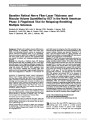
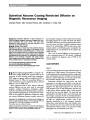
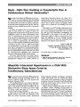
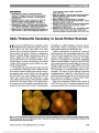
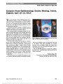
Show Baseline Retinal Nerve Fiber Layer Thickness and Macular Volume Quantified by OCT in the North American Phase 3 Fingolimod Trial for Relapsing-Remitting Multiple Sclerosis Kimberly M. Winges, MD, John S. Werner, PhD, Danielle J. Harvey, PhD, Kimberly E. Cello, BS, Mary K. Durbin, PhD, Laura J. Balcer, MD, MSCE, Peter A. Calabresi, MD, John L. Keltner, MD Background: Patients with multiple sclerosis (MS) demon-strate thinning of peripapillary retinal nerve fiber layer (RNFL) and decreased macular volume as measured by optical coherence tomography (OCT). To our knowledge, there are no previous reports from a large MS OCT database with strict quality control measures that quantitate RNFL and macula in patients with relapsing-remitting multiple sclerosis. Methods: The University of California Davis OCT Reading Center gathered OCT data at baseline as part of the North American phase 3 trial of fingolimod (Gilenya). Average RNFL thickness (RNFLT) and macular volume (TMV) were measured using time domain OCT (TD-OCT). RNFL quadrants, clock hours, and macular subfields were included. With strict quality control and accounting for signal strength differences, scans were categorized as "reduced" or "not reduced" for each field, based on being less than 5th percentile for age-matched controls derived from the normative database in the scanner software. Patients were deemed "abnormal" if at least 1 eye had reduced values for a given parameter. Pa-tients with abnormalities in corresponding RNFL and macular subfields were compared by cross-tabulation. Results: The TD-OCT data were prospectively collected from 939 of the 1,083 trial patients, 712 of whom met all final quality and data inclusion criteria. Of the final cohort, 242 (34.0%) demonstrated reduced (less than 5th percentile) average RNFLT in at least 1 eye. One hundred seventy-eight (25.0%) patients had reduced TMV. One hundred twenty-eight (18.0%) demonstrated both reduced TMV and RNFLT in the same eye, whereas 42 (5.8%) had reduced TMV and RNFLT in both eyes. Of the 242 patients with reduced average RNFL thickness, 128 (52.9%) also had reduced TMV. Fifty patients had reduced TMV in the absence of reduced RNFLT in at least 1 eye, a cohort prevalence of 7.0%. Quadrant and subfield analysis showed a predomi-nance of temporal and inferior RNFL thinning, with inferior macular thinning corresponding best to RNFL thinning. Conclusion: RNFL and macular thinning/volume loss is common at baseline in relapsing-remitting multiple sclerosis, as measured by TD-OCT. When the RNFL is thin, the macular volume is reduced in more than half of the patients. There is a population of reduced TMV without any reduction in RNFLT. Documenting the prevalence and distribution of these struc-tural abnormalities supports recent reports and suggests new retinal areas to probe for functional vision changes in MS. Journal of Neuro-Ophthalmology 2013;33:322-329 doi: 10.1097/WNO.0b013e31829c51f7 © 2013 by North American Neuro-Ophthalmology Society Retinal nerve fiber layer (RNFL) degeneration and the resulting optic nerve atrophy is a widely accepted measure of disease burden in patients with multiple sclerosis (MS) (1,2). It is thought that both anterograde Department of Ophthalmology (KMW), Veterans Administration Medical Center, Portland, Oregon; Department of Ophthalmology and Vision Science (KMW, JSW, KEC, JLK), Division of Biostatistics (DJH), Department of Public Health Sciences, and Departments of Neurology and Neurosurgery (JLK), University of California Davis School of Medicine, Davis, California; Carl Zeiss Meditec, Inc (MKD), Dublin, California; Department of Neurology (LJB), New York Uni-versity Langone Medical Center, New York, New York; and Department of Neurology (PAC), Johns Hopkins University School of Medicine, Baltimore, Maryland. Novartis Pharmaceuticals Corporation funded OCT data collection as part of the fingolimod phase 3 clinical trial NCT00355134. Partial support was also provided by an unrestricted grant from Research to Prevent Blindness to University of California, Davis, CA. KMW received support from the Department of Veterans Affairs. The authors report no conflicts of interest. Supplemental digital content is available for this article. Direct URL citations appear in the printed text and are provided in the full text and PDF versions of this article on the journal's Web site (www. jneuro-ophthalmology.com). Address correspondence to Kimberly M. Winges, MD, Department of Ophthalmology, Portland VA Medical Center, Mail Code P3EYE, 3710 SW US Veterans Hospital Road, Portland, OR, 97239; E-mail: kim.winges@gmail.com 322 Winges et al: J Neuro-Ophthalmol 2013; 33: 322-329 Original Contribution Copyright © North American Neuro-Ophthalmology Society. Unauthorized reproduction of this article is prohibited. and retrograde transsynaptic axon degeneration may be responsible for the loss of neural tissue in the brain and the eye affecting both the white and gray matter (3). It is evident both in magnetic resonance imaging lesion burden and on pathological specimens showing gliosis and neuronal loss, in addition to retinal ganglion cell axon degeneration (4-7). Optical coherence tomography (OCT) has gained increas-ing popularity in quantifying RNFL thickness (RNFLT) as a measure of axon disease in the MS population. Both time domain and spectral domain platforms are reproducible and reliable in quantifying these changes (1,8-12). Furthermore, peripapillary RNFL thinning correlates over time with clinical measures of low-contrast letter acuity and contrast sensitivity, as well as the expanded disability score and disease duration, giving clinicians an objective way to follow disease burden (9,12-15). Even patients with MS without a history of optic neuritis have shown thinner RNFL than controls (16,17), providing evidence that at baseline, patients with MS have abnormal optic nerves. It is now recognized that macular volume is reduced in MS vs normal eyes, and some studies have shown that macular volume loss is associated with RNFL loss (18,19). Approximately 34% of the macular volume is made up of ganglion cells and their axons, so it may be expected that macular volume loss would follow RNFL loss (8). However, OCT evidence of macular thinning has recently been dem-onstrated even in the absence of RNFL thinning, with new evidence of inner and outer macular atrophy (20). Thus, in early MS, there are significant fundamental structural changes of the retina that can be quantified in vivo. The purpose of our study was to use the largest known quality-controlled database of time domain OCT (TD-OCT) in a phase 3 MS trial to describe and map the baseline thickness and/or volume of the RNFL and macula in the relapsing-remitting MS population. METHODS In this retrospective observational study, OCT data were collected from all screening TD-OCT scans performed for FREEDOMS 2, the phase 3 North American trial of fingolimod (Gilenya), a sphingosine 1-phosphate receptor modulator that is the first Food and Drug Administration- approved oral treatment in the relapsing-remitting MS pop-ulation (21,22). Institutional review board approval was obtained at University of California Davis for this substudy. Patients Patients were recruited for FREEDOMS 2 based on the following inclusion criteria: men or nonpregnant women, 18-55 years of age, a diagnosis of MS as defined by 2005 revised McDonald criteria, a relapsing-remitting course with at least 1 documented relapse during the previous 1 year or 2 documented relapses during the 2 years before randomization, and an expanded disability status scale score of 0-5.5 inclusive (Novartis Protocol for North American Phase 3 Fingolimod Clinical Trial NCT00355134, 2006). During randomization, those patients in whom a suspicion of macular edema by dilated ophthalmoscopy or OCT (increased central foveal thickness or cystic changes in the fovea) failed screening and were not randomized into the study. Optical Coherence Tomography OCT scans were collected at the time of randomization and in follow-up over the 2-year study, using a single alignment and capture on the time domain platform (Stratus OCT; Carl Zeiss Meditec, Inc, Dublin, CA). Fast RNFLT protocols measured A-scans in a nominal 1.73-mm radius circle of peripapillary RNFL. Data for average RNFLT, 4 quadrants, and 12 clock hours were collected. The left eye from a sample MS patient with reduced RNFLT is shown in Figure 1A. Total macular volume (TMV) and the 9 Early Treatment of Diabetic Retinopathy Study (ETDRS) sectors were also collected, using fast macular thickness protocols to measure A-scans over the 1-mm central fovea, 4 quadrants of the 1- to 3-mm inner macular ring, and 4 quadrants of the 3- to 6-mm outer macular ring (23). The sameMS patient as in Figure 1A is shown in Figure 1B with reduced macular thickness. Data from both eyes of each subject were submitted to the study sponsor and to a centralized review by masked investigators at the University of California Davis OCT Reading Center. As published previously, scans were excluded from the final database if they met any of the following criteria: signal strength less than 7 (except in the case of a few clearly visible fovea or optic nerve scans with correct centration), exported data missing, extra scans, decentered scans, wrong scans or scanner used, or they required redraw by the technician due to segmentation artifact (24). Only patients with complete data for both eyes were included in the final OCT analysis. Measurements for each eye were categorized as "reduced" (less than 5th percentile of normal limits) or "normal, not reduced" (within or above normal limits) based on the manufacturer's normative data-base of age-matched controls (25). Study analysts were masked to all clinical information beyond gender, date of birth, and eye that was measured. Thus, the number of patients with a history of optic neuritis or glaucoma was unknown. Analysis Percentages of age-matched individuals who had abnor-mal results in a given field were graphed and summ-arized (See Supplemental Digital Content, Figure 1, http://links.lww.com/WNO/A81). Scans from reduced and normal eyes were also plotted according to measured field of interest (See Supplemental Digital Content, Figure 2, http://links.lww.com/WNO/A82, and Figure 3, http://links.lww.com/WNO/A83). Average OCT Winges et al: J Neuro-Ophthalmol 2013; 33: 322-329 323 Original Contribution Copyright © North American Neuro-Ophthalmology Society. Unauthorized reproduction of this article is prohibited. thicknesses were measured in micrometers ± one standard deviation (SD; type 1 error rate set at 0.05). Based on retinal ganglion cell axon distribution in the fundus, cross-tabulations of anatomically corresponding RNFL and macula quadrants were created. McNemar test was used to compare proportions of patients who had thinning in an RNFL quadrant and the corresponding outer or inner sector of the macula. Linear repeated-measures FIG. 1. Left eye of a patient with relapsing-remitting multiple sclerosis. A. Fast retinal nerve fiber layer thickness (RNFLT) protocol shows reduced RNFLT. B. Fast macular thickness protocol reveals reduced total macular volume and macular sector thickness. ETDRS, Early Treatment of Diabetic Retinopathy Study; OCT, optical coherence tomography; OS, left eye; TMV, total macular volume. 324 Winges et al: J Neuro-Ophthalmol 2013; 33: 322-329 Original Contribution Copyright © North American Neuro-Ophthalmology Society. Unauthorized reproduction of this article is prohibited. regression models, assuming an exchangeable correlation structure to account for the intercorrelation between eyes from the same person, were used to compare signal strength and mean thicknesses between reduced eyes and not reduced eyes (26). Models for mean thicknesses included signal strength as a covariate to ensure that differences were not due to differences in signal strength. RESULTS One thousand eighty-three patients were enrolled in the clinical trial. The OCT Reading Center received 18,733 scans from 939 patients at 96 sites. From this database, 2,880 high-quality representative OCT scans determined by the above strict quality control criteria (24) were selected. Only patients with complete data of each eye's macula and RNFL were used, representing 1,434 eyes in 717 patients. Across the 1,434 eyes, the average signal strength was 8.1 ± 1.5 for RNFL and 7.7 ± 1.5 for TMV. During final analysis, total average RNFLT and TMV were missing in five patients, but all subfield measures for these patients were included. Average RNFL Thickness vs Total Macular Volume Table 1 summarizes results of total average RNFLT and TMV. Of 712 patients, 242 (34.0%) had reduced average RNFLT. TMV was reduced in 178 (25.0%) patients. Average RNFLT and TMV were reduced in the same eye of 128 (18.0%) patients. Of the 242 patients with reduced average RNFLT, 128 (52.9%) also demonstrated reduced TMV. Fifty patients had reduced TMV without reduction in RNFLT in the same eye, representing 7.0% of all patients in the study cohort and 10.6% of all patients with a normal RNFLT. Of the 712 patients with average RNFLT and TMV available for both eyes, 153 (21.5%) demonstrated bilater-ally reduced values for one or both categories: bilateral RNFLT in 115 individuals, bilateral TMV in 80 individ-uals, and bilateral RNFLT and TMV in 42 patients. Twenty-one patients had bilaterally reduced TMV without RNFLT reduction in either eye. RNFL and Macular Subfield Thickness Figure 1A (See Supplemental Digital Content, http://links.lww.com/WNO/A81) summarizes the percent of patients with reduced RNFLT in at least one eye by quadrants and clock hours. RNFLT was most often reduced in the temporal quadrant (34.3% of the patients) and in clock hours 2 (41.7%) and 7 (52.7%). Reduced eyes had significantly lower signal strength for all RNFL measures (Table 2), but even after accounting for these differences, the average RNFL values were significantly different between normal and thin eyes (Table 3, P , 0.001 for all RNFL measures). Figure 1B (See Supplemental Digital Content, http://links. lww.com/WNO/A81) summarizes the percent of patients with reduced macular thickness in at least one eye by ETDRS subfield. TMV was 5.97 ± 0.20 mm3 for normal eyes and 6.83 ± 0.38 mm3 for reduced eyes. In general, the frequency of reduced thickness in the inner quadrants was higher than that in the outer quadrants. The highest frequency of macular thinning was found in the inner temporal (37.4%) and inner inferior (34.9%) subfields. The lowest frequency regions of thinning were the central 1-mm fovea (7.8%) and the outer nasal quadrant (2.5%). Reduced eyes had significantly lower signal strength for TMV and the outer quadrants (Table 4), but even after accounting for these differences, all macular values were significantly different between normal and thin eyes (Table 5; P , 0.001 for all macular measures). RNFL quadrants and macular sectors that corre-sponded anatomically are compared in Table 6. The per-centage of patients with thinning in the RNFL quadrant that were also thin in the macula quadrant is also pre-sented. The highest frequency of concurrent thinning in RNFL and macula quadrants occurred in the superior RNFL and inner temporal macula (17.1%). Thinning of the inferior RNFL was associated with the inner tem-poral macula (16.9%), inner inferior macula (16.5%), and outer inferior macula (15.5%). In most cases, per-centages between inner and outer macula quadrant and RNFLT were significantly different (P , 0.001, McNemar test). DISCUSSION Our study demonstrated baseline thinning of the RNFL reduced macular volume in the largest quality-controlled data set of OCT from a phase 3 MS trial cohort to date. In this population, about one third of the patients had RNFL thinning at baseline and one-quarter of patients had reduced TMV. Average RNFLT and TMV were collec-tively reduced in 18.0%. When the RNFLT was reduced, the macular volume was reduced in over half of the patients (52.9%). In other words, individuals with RNFL reduction were much more likely to have TMV reduction TABLE 1. Patients with abnormal TMV vs abnormal average RNFLT in at least one eye RNFLT Normal, n (%) RNFLT Abnormal, n (%) TMV normal 420 (59.0) 114 (16.0) 534 (75.0) TMV abnormal 50 (7.0) 128 (18.0) 178 (25.0) Total 470 (66.0) 242 (34.0) 712 RNFLT, retinal nerve layer thickness; TMV, total macular volume. Winges et al: J Neuro-Ophthalmol 2013; 33: 322-329 325 Original Contribution Copyright © North American Neuro-Ophthalmology Society. Unauthorized reproduction of this article is prohibited. (128 of 242 patients, 52.9%) than individuals without RNFL reduction (50 of 470 patients, 10.6%). In total, macular volume was reduced in the absence of RNFL reduction in 7.0% of the trial cohort. Additionally, many subfields of RNFL and macula were abnormal, with the inferior RNFL and inferior and temporal macula showing the most collectively reduced values. There is no doubt that a significant fraction of this cohort had objective TABLE 2. Signal strength differences in reduced and not reduced eyes for retinal nerve fiber layer thickness RNFL Region Average Signal Strength of Reduced Eyes (SD) Average Signal Strength of Not Reduced Eyes (SD)* P Average thickness RNFL 7.75 (1.54) 8.31 (1.44) ,0.001 Superior average RNFL 7.86 (1.52) 8.25 (1.46) ,0.001 Inferior average RNFL 7.71 (1.51) 8.29 (1.45) ,0.001 Temporal average RNFL 7.79 (1.56) 8.27 (1.45) ,0.001 Nasal average RNFL 7.78 (1.45) 8.23 (1.49) ,0.001 Clock hour 1† 7.79 (1.59) 8.27 (1.45) ,0.001 Clock hour 2 7.89 (1.48) 8.27 (1.49) ,0.001 Clock hour 3 7.75 (1.51) 8.30 (1.45) ,0.001 Clock hour 4 7.76 (1.62) 8.27 (1.44) ,0.001 Clock hour 5 7.69 (1.56) 8.23 (1.48) ,0.001 Clock hour 6 7.76 (1.50) 8.24 (1.47) ,0.001 Clock hour 7 7.93 (1.50) 8.25 (1.49) 0.007 Clock hour 8 7.73 (1.58) 8.29 (1.44) ,0.001 Clock hour 9 7.75 (1.57) 8.26 (1.45) ,0.001 Clock hour 10 7.78 (1.57) 8.25 (1.46) ,0.001 Clock hour 11 7.70 (1.57) 8.30 (1.44) ,0.001 Clock hour 12 7.89 (1.54) 8.20 (1.47) 0.009 *Includes all normal eyes of individuals with both eyes not reduced. †Clock hours are standardized such that left eyes have been flipped to correspond with right eye orientation, that is, 9-o'clock position corresponds to the temporal quadrant and 3-o'clock position corresponds to the nasal quadrant. RNFL, retinal nerve fiber layer; SD, standard deviation. TABLE 3. Distribution of RNFL thinning by quadrant and clock hour in relapsing-remitting multiple sclerosis patients with at least one eye affected RNFL Region Percent of Patients With at Least One Eye ,5% Normal Thickness, % Average Value of Reduced Eyes, mm (SD) Average Value of Not Reduced Eyes, mm (SD)* Average thickness RNFL 34.0† 73.3 (8.2) 100.1 (9.9) Superior average RNFL 28.3 86.2 (11.1) 123.9 (15.2) Inferior average RNFL 26.6 85.4 (10.9) 126.2 (16.6) Temporal average RNFL 34.3 41.1 (6.1) 68.4 (11.8) Nasal average RNFL 17.6 46.8 (6.2) 81.1 (17.0) Clock hour 1‡ 27.6 70.3 (11.7) 118.8 (21.1) Clock hour 2 41.7 51.7 (10.2) 92.0 (19.7) Clock hour 3 36.4 34.3 (5.1) 61.4 (16.0) Clock hour 4 29.8 40.7 (6.9) 75.3 (17.6) Clock hour 5 15.6 60.3 (9.5) 117.7 (25.9) Clock hour 6 21.8 79.0 (13.8) 136.5 (23.8) Clock hour 7 52.7 78.3 (14.0) 127.6 (20.7) Clock hour 8 27.7 40.2 (5.9) 73.8 (17.1) Clock hour 9 27.6 32.6 (4.0) 57.7 (14.7) Clock hour 10 24.5 46.5 (7.8) 87.5 (20.6) Clock hour 11 32.5 80.5 (13.8) 126.3 (19.6) Clock hour 12 21.1 73.2 (11.7) 126.3 (22.4) *Includes all normal eyes of individuals with both eyes not reduced. †Average RNFL% is out of 712 patients due to 5 patients missing only this measurement. All sectors and clock hours are percentage out of 717 patients. ‡Clock hours are standardized such that left eyes have been flipped to correspond with right eye orientation, that is, 9-o'clock position corresponds to the temporal quadrant and 3-o'clock position corresponds to the nasal quadrant. RNFL, retinal nerve fiber layer; SD, standard deviation. 326 Winges et al: J Neuro-Ophthalmol 2013; 33: 322-329 Original Contribution Copyright © North American Neuro-Ophthalmology Society. Unauthorized reproduction of this article is prohibited. OCT evidence of structural damage to not only the optic nerve but the macula as well at baseline. The fact that RNFL and macular thinning are linked in this population is not a new idea (8,17,19). However, this study is arguably the largest database of the MS OCT trial data that has undergone strict quality control with a cen-tralized OCT reading center. Additionally, this study detailed which subfields of the macula and quadrants or clock hours of the optic nerve were preferentially affected. They were often affected together, particularly in the inferior region, likely representing inferior peripapil-lary RNFL from ganglion cells with axons originating in the inferior macula. Our study also supported the notion that a subset of patients with MS may have structural damage to the macula in the absence of damage to the RNFL. Saidha et al (20) described the preferential macular involvement in 10% of their 450 patients with MS as measured by the spectral domain OCT, who appeared to have worse disease severity scores and more severe macular function by multifocal electroretinography. Our results showed that 7.0% of 712 patients (10.6% of the patients with a normal RNFLT) had reduced TMV in the absence of thin average RNFL. Our data collected with TD-OCT supported the findings of Saidha et al. Reduced central foveal area in 7.8% of our patients also demonstrated that MS can affect the outer retinal layers. This has been observed in patients with long-standing MS using adaptive optics (27,28). Patients with a history of MS tend to develop peripapil-lary RNFL thinning in the temporal quadrant, which preferentially affects the papillomacular bundle (29). In our study, the papillomacular bundle region showed great thickness variability. Thinning of the temporal optic nerve quadrant occurred in 246 patients (34.3%). The inner nasal macula was thin in 20.8%, and the outer nasal macula was thin in only 2.5%. The inner macular ring encompasses a thicker ganglion cell layer than that of the outer macular ring (30), which may offer a possible explanation for this disparity because ganglion cells in the inner sector would contribute proportionally more to overall macular thickness TABLE 4. Signal strength differences in reduced and not reduced eyes for macula Macular Subfield Average Signal Strength of Reduced Eyes (SD) Average Signal Strength of Not Reduced Eyes (SD)* P Total macular volume 7.44 (1.53) 7.79 (1.51) 0.01 Outer superior average thickness 7.30 (1.42) 7.78 (1.53) 0.003 Outer nasal average thickness 7.09 (1.37) 7.75 (1.53) 0.04 Outer inferior average thickness 7.40 (1.52) 7.82 (1.50) ,0.001 Outer temporal average thickness 7.43 (1.53) 7.84 (1.51) ,0.001 Inner superior average thickness 7.61 (1.49) 7.76 (1.52) 0.24 Inner nasal average thickness 7.61 (1.50) 7.76 (1.53) 0.28 Inner inferior average thickness 7.59 (1.54) 7.76 (1.50) 0.21 Inner temporal average thickness 7.68 (1.56) 7.74 (1.51) 0.52 Central foveal area thickness 7.84 (1.59) 7.72 (1.53) 0.49 *Includes all normal eyes of individuals with both eyes not reduced. TABLE 5. Distribution of macular thickness by subfield in relapsing-remitting multiple sclerosis patients with at least one eye affected Macular Subfield Percent of Patients With at Least One Eye ,5% Normal, % Average Value of Reduced Eyes, mm (SD)* Average Value of Not Reduced Eyes, mm (SD)† TMV 25.3‡ 5.97 (0.20) 6.83 (0.38) Outer superior average thickness 14.1 200.8 (6.9) 234.5 (14.9) Outer nasal average thickness 2.5 193.3 (3.8) 245.8 (19.9) Outer inferior average thickness 28.4 199.0 (7.7) 230.8 (13.8) Outer temporal average thickness 28.3 193.8 (6.8) 221.4 (15.5) Inner superior average thickness 23.7 233.5 (9.5) 270.8 (15.1) Inner nasal average thickness 20.8 229.2 (10.5) 269.6 (16.9) Inner inferior average thickness 34.9 236.0 (12.3) 271.0 (18.0) Inner temporal average thickness 37.4 228.9 (9.8) 261.1 (17.1) Central foveal area thickness 7.8 159.6 (7.3) 203.6 (21.6) *Except for TMV, which is measured in cubic millimeters. †Includes all normal eyes of individuals with both eyes not reduced. ‡TMV% is of 712 patients due to 5 patients missing only this measurement. All sectors are percent of 717 patients. TMV, total macular volume. Winges et al: J Neuro-Ophthalmol 2013; 33: 322-329 327 Original Contribution Copyright © North American Neuro-Ophthalmology Society. Unauthorized reproduction of this article is prohibited. than other retinal layers. Nevertheless, other macular sectors did not demonstrate such a dramatic difference. Although the temporal RNFL was the most commonly thinned quadrant, as reported in previous studies (29), the clock hours of greatest thinning were in the inferior quad-rant. Since the inferior RNFL is traditionally the thickest quadrant, it is not surprising that the inferior RNFL would show the most thinning since there are more nerve fibers at risk. A true comparative subfield analysis was limited by predetermined normative database and analysis plots for ETDRS sectors in the macula and RNFL quadrants. These regions may not optimally correspond in anatomical distribution. For example, the superior and inferior RNFL both represent axons from the temporal macula. Also, nominal categories of reduced and not reduced did not allow for continuous variable analysis to determine which patients had values near but not beyond the threshold of 5% (i.e., low-normal, just above 5%), the analysis of which may clarify any linear relationships between RNFL and macular thinning. The lack of segmentation algorithms in TD-OCT precludes further interpretation of our data set. As technol-ogy improves and time domain platforms give way to spectral domain and high-resolution OCT, intraretinal OCT segmentation algorithms and volume mapping are now beginning to detail how much of macular thinning is due to ganglion cell death or RNFL loss vs damage to outer retinal layers (20,27,28,31,32). In conclusion, our study showed that both the RNFL and macula are commonly thinned in the relapsing- remitting MS population at baseline. We confirmed that when the average RNFL is thin, the macula showed reduced volume at baseline. We also documented a population of patients in whom the macula was preferentially affected, despite normal RNFL as measured by TD-OCT. Future assessment of the longitudinal and clinical data from the MS fingolimod trial should provide insights into the pop-ulations at risk of macular pathology and loss of visual function. ACKNOWLEDGMENTS The authors would like to thank Novartis employees Clinical Trial Head Neuroscience Tracy Stites, Head of Neuroimmunology Clinical Science Unit Francis Gordon, and Global Program Medical Director Philipp von Rose-nstiel for their help in data acquisition and management. The authors would also like to thank Patricia Duffel and Dustin McGranahan at the University of Iowa Department of Ophthalmology for logistical help in preparation of this article. REFERENCES 1. Kardon RH. Role of the macular optical coherence tomography scan in neuro-ophthalmology. J Neuroophthalmol. 2011;31:353-361. 2. Rudick RA, Lee JC, Nakamura K, Fisher E. Gray matter atrophy correlates with MS disability progression measured with MSFC but not EDSS. J Neurol Sci. 2009;282:106-111. 3. Petzold A, de Boer JF, Schippling S, Vermersch P, Kardon R, Green A, Calabresi PA, Polman C. Optical coherence tomography in multiple sclerosis: a systematic review and meta-analysis. Lancet Neurol. 2010;9:921-932. 4. Trip SA, Schlottmann PG, Jones SJ, Li WY, Garway-Heath DF, Thompson AJ, Plant GT, Miller DH. Optic nerve atrophy and retinal nerve fibre layer thinning following optic neuritis: evidence that axonal loss is a substrate of MRI-detected atrophy. Neuroimage. 2006;31:286-293. 5. Frisén L, Hoyt WF. Insidious atrophy of retinal nerve fibers in multiple sclerosis. Funduscopic identification in patients with and without visual complaints. Arch Ophthalmol. 1974;92:91- 97. 6. Kerrison JB, Flynn T, Green WR. Retinal pathologic changes in multiple sclerosis. Retina. 1994;14:445-451. 7. Green AJ, McQuaid S, Hauser SL, Allen IV, Lyness R. Ocular pathology in multiple sclerosis: retinal atrophy and inflammation irrespective of disease duration. Brain. 2010;133:1591-1601. 8. Paunescu LA, Schuman JS, Price LL, Stark PC, Beaton S, Ishikawa H, Wollstein G, Fujimoto JG. Reproducibility of nerve fiber thickness, macular thickness, and optic nerve head measurements using StratusOCT. Invest Ophthalmol Vis Sci. 2004;45:1716-1724. 9. Burkholder BM, Osborne B, Loguidice MJ, Bisker E, Frohman TC, Conger A, Ratchford JN, Warner C, Markowitz CE, Jacobs DA, Galetta SL, Cutter GR, Maguire MG, Calabresi PA, Balcer LJ, Frohman EM. Macular volume determined by optical coherence tomography as a measure of neuronal loss in multiple sclerosis. Arch Neurol. 2009;66:1366-1372. 10. Walter SD, Ishikawa H, Galetta KM, Sakai RE, Feller DJ, Henderson SB, Wilson JA, Maguire MG, Galetta SL, Frohman E, Calabresi PA, Schuman JS, Balcer LJ. Ganglion cell loss in relation to visual disability in multiple sclerosis. Ophthalmology. 2012;119:1250-1257. 11. Syc SB, Warner CV, Hiremath GS, Farrell SK, Ratchford JN, Conger A, Frohman T, Cutter G, Balcer LJ, Frohman EM, TABLE 6. Comparison of reduced retinal nerve fiber layer thickness to reduced macular thickness by quadrant or sector RNFL Quadrant Macular Subfield Percent of Patients With Reduced RNFLT and Macular Thickness in the Same Eye, % Superior Outer superior 7.9 Superior Inner superior 11.4 Superior Outer temporal 12.4 Superior Inner temporal 17.1 Inferior Outer inferior 15.5 Inferior Inner inferior 16.5 Inferior Outer temporal 13.7 Inferior Inner temporal 16.9 Temporal Outer nasal 2.1 Temporal Inner nasal 11.8 RNFL average Total macular volume 17.8 RNFL average Central 1 mm thickness 3.8 RNFL, retinal nerve fiber layer; RNFLT, retinal nerve fiber layer thickness. 328 Winges et al: J Neuro-Ophthalmol 2013; 33: 322-329 Original Contribution Copyright © North American Neuro-Ophthalmology Society. Unauthorized reproduction of this article is prohibited. Calabresi PA. Reproducibility of high-resolution optic coherence tomography in multiple sclerosis. Mult Scler. 2010;16:827- 839. 12. Kallenbach K, Frederiksen J. Optical coherence tomography in optic neuritis and multiple sclerosis: a review. Eur J Neurol. 2007;14:841-849. 13. Fisher JB, Jacobs DA, Markowitz CE, Galetta SL, Volpe NJ, Nano-Schiavi ML, Baier ML, Frohman EM, Winslow H, Frohman TC, Calabresi PA, Maguire MG, Cutter GR, Balcer LJ. Relation of visual function in retinal nerve fiber layer thickness in multiple sclerosis. Ophthalmology. 2006;113:324-332. 14. Sakai RE, Feller DJ, Galetta KM, Galetta SL, Balcer LJ. Vision in multiple sclerosis: the story, structure-function correlations, and models for neuroprotection. J Neuroophthalmol. 2011;31:362-373. 15. Greenberg B, Frohman E. Optical coherence tomography as a potential readout in clinical trials. Ther Adv Neurol Disord. 2010;3:153-160. 16. Pueyo V, Ara JR, Almarcegui C, Martin J, Güerri N, García E, Pablo LE, Honrubia FM, Fernandez FJ. Sub-clinical atrophy of the retinal nerve fibre layer in multiple sclerosis. Acta Ophthalmol. 2010;88:748-752. 17. Talman LS, Bisker ER, Sackel DJ, Long DA Jr, Galetta KM, Ratchford JN, Lile DJ, Farrell SK, Loguidice MJ, Remington G, Conger A, Frohman TC, Jacobs DA, Markowitz CE, Cutter GR, Ying GS, Dai Y, Maguire MG, Galetta SL, Frohman EM, Calabresi PA, Balcer LJ. Longitudinal study of vision and retinal nerve fiber layer thickness in multiple sclerosis. Ann Neurol. 2010;67:749-760. 18. Trip SA, Schlottmann PG, Jones SJ, Altmann DR, Garway- Heath DF, Thompson AJ, Plant GT, Miller DH. Retinal nerve fiber layer axonal loss and visual dysfunction in optic neuritis. Ann Neurol. 2005;58:383-391. 19. Watson GM, Keltner JL, Chin EK, Harvey D, Nguyen A, Park SS. Comparison of retinal nerve fiber layer and central macular thickness measurements among five different optical coherence tomography instruments in patients with multiple sclerosis and optic neuritis. J Neuroophthalmol. 2011;31:110-116. 20. Saidha S, Syc SB, Ibrahim MA, Eckstein C, Warner CV, Farrell SK, Oakley JD, Durbin MK, Meyer SA, Balcer LJ, Frohman EM, Rosenzweig JM, Newsome SD, Ratchford JN, Nguyen QD, Calabresi PA. Primary retinal pathology in multiple sclerosis as detected by optical coherence tomography. Brain. 2011;134:518-533. 21. Kappos L; FREEDOMS Study Group. A placebo-controlled trial of oral fingolimod in relapsing multiple sclerosis. New Engl J Med. 2010;362:387-401. 22. Cohen JA, Barkhof F, Giancarlo C, Hartung H-P, Khatri BO, Montalban X, Pelletier J, Capra R, Gallo P, Izquierdo G, Tiel-Wilck K, de Vera A, Jin J, Stites T, Wu S, Aradhye S, Kappos L; TRANSFORMS Study Group. Oral fingolimod or intramuscular interferon for relapsing multiple sclerosis. N Engl J Med. 2010;362:402-415. 23. Early Treatment Diabetic Retinopathy Study Research Group. Grading diabetic retinopathy from stereoscopic color fundus photographs-an extension of the modified Airlie House classification. ETDRS report number 10. Ophthalmology. 1991;98:786-806. 24. Keltner J, Cello KE, Balcer LJ, Calabresi PA, Markowitz CE, Werner JS. Stratus OCT quality control in two multi-center multiple sclerosis clinical trials. Neuroophthalmology. 2011;35:7-64. 25. Patella VM. STRATUSOCTTM: Establishment of Normative Reference Values for Retinal Nerve Fiber Layer Thickness Measurements. Dublin, CA: Carl Zeiss Meditec, Inc, 2003. 26. Diggle PJ, Liang KY, Zeger SL. Analysis of Longitudinal Data, 1st edition. Oxford, United Kingdom: Clarendon Press, 1994. 27. Werner JS, Keltner JL, Zawadzki RJ, Choi SS. Outer retinal abnormalities associated with inner retinal pathology in nonglaucomatous and glaucomatous optic neuropathies. Eye (Lond). 2011;25:279-289. 28. Choi SS, Zawadski RJ, Keltner JL, Werner JS. Changes in cellular structures revealed by ultra-high resolution retinal imaging in optic neuropathies. Invest Ophthalmol Vis Sci. 2008;49:2103-2119. 29. Bock M, Brandt AU, Dörr J, Kraft H, Weinges-Evers N, Gaede G, Pfueller CF, Herges K, Radbruch H, Ohlraun S, Bellmann- Strobl J, Kuchenbecker J, Zipp F, Paul F. Patterns of retinal nerve fiber layer loss in multiple sclerosis patients with or without optic neuritis and glaucoma patients. Clin Neurol Neurosurg. 2010;112:647-652. 30. Curcio CA, Allen KA. Topography of ganglion cells in human retina. J Comp Neurol. 1990;300:5-25. 31. Seigo MA, Sotirchos ES, Newsome S, Babiarz A, Eckstein C, Ford E, Oakley JD, Syc SB, Frohman TC, Ratchford JN, Balcer LJ, Frohman EM, Calabresi PA, Saidha S. In vivo assessment of retinal neuronal layers in multiple sclerosis with manual and automated optical coherence tomography segmentation techniques. J Neurol. 2012;259:2119-2130. 32. Davies EC, Galetta KM, Sackel DJ, Talman LS, Frohman EM, Calabresi PA, Galetta SL, Balcer LJ. Retinal ganglion cell layer volumetric assessment by spectral-domain optical coherence tomography in multiple sclerosis: Application of a high-precision manual estimation technique. J Neuroophthalmol. 2011;31:260-264. Winges et al: J Neuro-Ophthalmol 2013; 33: 322-329 329 Original Contribution Copyright © North American Neuro-Ophthalmology Society. Unauthorized reproduction of this article is prohibited. |