| OCR Text |
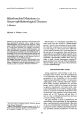
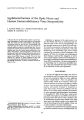
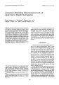
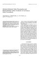
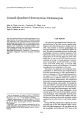
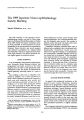
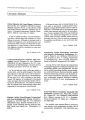
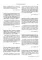
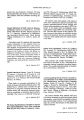
Show Journal of Clinical Neuro- ophthalmology 10( 3): 219- 222, 1990. © 1990 Raven Press, Ltd., New York Crossed- Quadrant Homonymous Hemianopsia John A. Dyer, M. B. B. S., Lawrence W. Hirst, M. D., Kevin Vandeleur, ( Snr) M. B. B. S., Terence Carey, M. B. B. S., and Peter R. Mann M. B. B. S. This case report illustrates the clinicopathological correlation between the anatomic defect of bilateral occipital lobe infarction with crossed- quadrant homonymous hemianopsia that resulted from a cervical spine injury. Routine ophthalmological and neuroradiological investigations were undertaken, including magnetic resonance imaging scanning, that demonstrate some important characteristics of this rare field defect. Key Words: Hemianopsia- Perimetry- Computered tomography- Magnetic resonance imaging- Occipital lobe. From the Department of Surgery, Division of Ophthal", lology, a. A. D., L. W. H.) Princess Alexandra Hospital, and Wickham Terrace, ( K. V., T. C., P. R. M.) Brisbane, Australia. Address correspondence and reprint request to Profes~ or Lawrence W. Hirst, 2nd Floor, Lions Clinical Research Building, Ipswich Road, Woolloongabba, Brisbane, Queensland 4102, Australia. Supported in part by the Australian Foundation f~ r the Prevention of Blindness ( Queensland Division), The Pnncess Alexandra Hospital, and the Royal Brisbane Hospital, Brisbane, Australia. 219 CASE REPORT A 30- year- old, right- handed man suffered a fracture- dislocation of his fifth cervical vertebra after hitting a log while diving into a shallow pool in 1960. This resulted in transient quadriplegia and transient blindness for an undisclosed period of time. He made a remarkable recovery of vision and quadriplegia. However, residual weakness of his right hand and leg remained, with loss of pain and temperature on his left side below the level of the nipple line. It was not until 1975 that he was documented as having a crossed- quadrant homonymous hemianopsia visual field defect on Goldmann visual field testing ( Fig. lA and B) while undergoing a routine eye examination. No further management was undertaken at this time. The patient was diagnosed as having type II hyperlipidemia in 1978 and his past surgical history included appendectomy in 1938, transurethral resection of prostate in 1978, and right inguinal hernia repair in 1979. In 1985 examination showed corrected vision of 20/ 20 for both eyes, normal intraocular pressure measurement, and visual field testing of a crossed- quadrant hemianopsia. In June 1986, while reversing a boat trailer, he twisted his neck and suffered a syncopal attack and total loss of power to the right hand. He was reviewed by both a neurologist and neurosurgeon and underwent investigation with plain radiography of the cervical spine, CAT scan of cervical spine, and cervical myelogram. The radiographs showed previous fracture of the fifth cervical vertebra with partial bony ankylosis to the fourth and sixth cervical vertebra but no other bony or soft tissue pathology. In May 1987 the patient had an episode of severe transient pain in the left temporal region associated with right hemiparesis. He was again seen by his neurologist and underwent CAT scan of the 220 J. A. DYER ET AL. FIG. 1. A: Goldmann visual field of left eye. B: Goldmann visual field of right eye demonstrating crossed- quadrant homonymous hemianopsia with central isthmus of intact vision. 1: Io- r-•• 1! riI1._ ,.. ." t>, -. ./ / ~ -- •• 1 · . " I I .... . It-- ::;' I -- ..,. " n .", \--- I •.• ': .~' ..' 1A 1! HIUflll head and a carotid duplex scan. The CAT scan ( Fig. 2) demonstrated a low- density region in the left occipital lobe consistent with old cerebral infarct. The carotid duplex scan was within normal limits. In August 1987 the patient was referred for further ophthalmological evaluation. Examination at that time showed corrected visual acuity 20/ 20 in each eye and reading vision of N4.5 in the right eye and N5.0 in the left eye for near. Pupil reactions were normal with no evidence of an afferent pupil defect. Extraocular motility was full and optokinetic nystagmus testing was normal in all directions. Corneal sensation was equal in both eyes. Intraocular pressure measurement by pneumotonometry was 15 mm Hg for both eyes. He had evidence of an early anterior supcapsular cataract change in the left lens. The fundus examination was normal. Humphrey's computer field analysis ( Fig. 3A and B) demonstrated crossed- quadrant homonymous field defect. On November 23, 1987, the patient underwent a magnetic resonance imaging ( MRI) scan ( Fig. 4), which demonstrated both right and left occipital pole disease with the lesions on the left side being more extensive than those on the right. DISCUSSION Cases of crossed- quadrant homonymous hemianopsia are rare. The most recent article ( 1) reviewed nine cases of this condition, documented worldwide from 1891 to 1982. Although the reports of these cases were incomplete in areas due to data availability, the authors noted some important characteristics of this visual field defect. / Gin Neuro- ophllwlmol, Vol. 10, No. 3, 1990 CROSSED- QUADRANT HOMONYMOUS HEMIANOPSIA 221 H. r · ! r. '--':" 1 8 3.1. [ 1': d". 11 ", II • I ... ,. l- • ' I ~. I .,.... j. ''" .11.... 1' ." I :. ", I '-- ,... • _ ~ "'. 11 A. I,', • , ' 1;" I. I I · .- " · ~, r- J ••",. r:- ,,' 1. ., I" L ..... I ,,... I I I to . pr • IC' I ,- ", I'~ II rIO' ... I ,', I Ir • I " " J". r I '-, 11 ~ I., I' /. I FIG. 2. CAT scan. Note low density region in left occipital lobe consistent with old cerebral infarct ( arrow). FIG. 3. A: Humphrey's computer field analyzer left eye. B: Humphrey's computer field analyzer right eye. Crossed- quadrant homonymous field defect is demonstrated in both eyes. sentation. The most frequent cause was embolization of the calcarine arteries ( from basilar or vertebral branches), or thromboembolism from associated cardiac disorders. 1. The defects may arise in ( a) two simultaneous quadrantanopsias; ( b) two successive homonymous hemianopsias, each resolving into a quadrantanopsia; or ( c) simultaneous bilateral homonymous hemianopsias resolving into crossedquadrant defects. 2. The defects usually extend across the horizontal meridian but respect the vertical midline. 3. There may be sparing of the monocular temporal crescent. 4. Central vision is usually preserved with a small isthmus of unaffected field connecting the two intact quadrants. 5. The intact quadrants may demonstrate small areas of field loss. 6. Color vision may be abnormal. 7. Riddoch phenomenon may be present, accounting for an apparent incongruity of field testing if a different velocity is used when testing isopters in different eyes. 8. Simple or complex visual hallucinations may appear in the blind or seeing field. Symonds and MacKenzie ( 2) have studied the origin of bilateral hemianopsias in some detail. They noted visual field loss was sudden or gradual and could be affected either simultaneously or in succession, with the former the more common pre- " I , . j " '. I-,! I" I .. L I, _~ I.. I'I"."~ 1 ~ • I )~ I i, -,- I "~.:..' • '- 1,__ I • I .11 I, • It .... J J Clin Neuro- ophtlullmol, Vol. 10, No. 3, 1990 222 ]. A. DYER ET AI. FIG. 4. Axial MRI scan. Demonstrating both right and left occipital pole disease ( arrows). J 0", t'. · The underlying etiology in this c . _ l~ most likely associated with vertebrobasilar disease or posterior cerebra! ischemia from the original cervical spine injury. Treatment of patients with this field defect is dependent on the extent of the defect. The use of both mirror or " checkerboard" prisms have been reported in the literature with varying degrees of success (~ 5). This case report illustrates many of the important characteristics discussed by Cross and Smith ( 1), and is the first case documented by computerized field analysis and MRI scanning. REFERENCES 1. Cross SA, Smith JL. Crossed- quadrant homonymous hemianopsia. The " checkerboard" field defect. / Clin Neuro Ophthaimol 1982; 2: 149- 58. 2. Symonds C, MacKenzie I. Bilateral loss of vision from cerebral infarction. Brain 1957; 80: 415- 55. 3. Walsh 11, Smith JL. Hemianopic spectacles. Am / Ophthalmol 1966; 61: 914- 5. 4. Mintz MJ. A mirror for hemianopia. Am I Ophthalmol 1979; 88: 768. 5. Smith JL, Weiner IG. Hemianopic fresnel prisms. / Clin Neuro OphthalmoI1982; 2: 19- 22. |