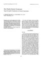
OCR Text |
Show Journal of Clinical Neuro- ophtluJlmology 10( 3): 167- 174, 1990. Neuro- ophthalmic Findings in Acquired Immunodeficiency Syndrome Ahmad M. Mansour, M. D. © 1990 Raven Press, Ltd., New York We reviewed the neuro- ophthalmic findings in 177 subjects with the acquired immunodeficiency syndrome ( AIDS) or AIDS- related complex who underwent an eye examination in one center from January 1984 to May 1989. The findings included ocular motor nerve palsies ( five cases), papilledema ( two cases), cytomegalovirus optic neuritis ( two cases), cortical blindness ( one case), conjugate gaze palsy ( one case), and altitudinal visual field defect ( one case). These findings were attributed to central nervous system toxoplasmosis ( four cases) or lymphoma ( one case), cryptococcal meningitis ( two cases), systemic cytomegalovirus infections ( two cases), and herpes simplex encephalitis ( one case). Of 177 patients, 61 patients were tested for syphilis. Twenty- six patients had positive rapid plasma reagin titers, and 28 had positive fluorescent treponemal antibody- absorbed tests. Human immunodeficiency virus- infected individuals need to be screened routinely for syphilis. Key Words: Acquired immunodeficiency syndromeCortical blindness- Cryptococcal meningitis- Cytomegalovirus optic neuritis- Gaze palsy- Herpes simplex encephalitis- Human immunodeficiency virusLymphoma- Ocular motor nerve palsy- Papilledema- Syphilis- Toxoplasmosis. From the Department of Ophthalmology, University of Texas Medical Branch, Galveston, Texas. Address correspondence and reprint requests to Ahmad Mansour, M. D., Ophthalmology- UTMB, Galveston, IX 77550, U. S. A. 167 The acquired immunodeficiency syndrome ( AIDS) is marked by the occurrence of various opportunistic infections and malignancies in previously healthy individuals. The most common ocular findings in subjects with AIDS include cottonwool spots and cytomegalovirus retinitis. The neuro- ophthalmic findings in one series of patients with AIDS are presented, MATERIALS AND METHODS We reviewed prospectively the ocular findings of patients with AIDS or AIDS- related complex examined by the author from September 1987 to May 1989, together with a retrospective review of subjects seen from January 1984 to August 1987 in the Department of Ophthalmology. The systemic findings of each patient were reviewed, focusing on opportunistic infections, malignancies, lymphocyte marker profile ( T4! f8 or T helperff suppressor ratio), and time interval from the neuro- ophthalmic findings to death. The majority of the patients had serum titers drawn against Toxoplasma gondii, herpes simplex, cytomegalovirus, and EpsteinBarr virus. Several patients were tested for syphilis using the fluorescent treponemal antibody absorbed test ( FTA- ABS), and the rapid plasma reagin ( RPR) test. RESULTS The results are presented in Tables 1 and 2. Of 177 patients, 127 had AIDS and 50 had AIDSrelated complex. None of the 50 patients with AIDS- related complex had neuro- ophthalmic complications. Ten of the 127 patients with AIDS had neuro- ophthalmic findings. These ten patients had a mean age of 34 years ( range 27-- 46), and consisted of nine men and 1 woman, with a racial distribution of six whites, two blacks, and two 168 AHMAD M. MANSOUR TABLE 1. Neuro- ophthalmic findings in 177 patients with AIDS or AIDS- related complex CNS, central nervous system. Cases reported are from the University of Texas Medical Branch, Galveston. Ocular motor nerve palsies Oculomotor nerve Trochlear nerve Abducens nerve Papilledema CNS toxoplasmosis Cryptococcal meningitis Cytomegalovirus optic neuritis Direct spread of cytomegalovirus retinitis Gaze palsy Paramedian pontine reticular formation lesion Cortical blindness Cryptococcal meningitis Visual field defect Inferior altitudinal field defect Multiple scotomas No. cases 1 ( CNS toxoplasmosis) 2 ( CNS toxoplasmosis and CNS lymphoma) 1 ( CNS toxoplasmosis) 2 1 ( CNS toxoplasmosis) 1 ( cryptococcal meningitis) 1 ( toxoplasmic encephalitis) Latin Americans. Neuro- ophthalmic findings included ocular motor nerve palsies ( five cases) ( Figs. 1- 4), papilledema ( two cases), and cytomegalovirus optic neuritis ( two cases) ( Figs. 5 and 6). Less common findings included cortical blindness ( one case), conjugate gaze palsy from a lesion of the paramedian pontine reticular formation ( one case), and an inferior altitudinal visual field defect ( one case) ( Fig. 7). The ocular findings in four cases were attributed to focal midbrain toxoplasmic lesions. The diagnosis of cerebral toxoplasmosis was established by the appearance of the lesions with and without contrast on computed tomography of the head, elevated serum antibody to Toxoplasma, a positive response to Toxoplasma chemotherapy, and finding of tachyzoites or tissue cysts on needle brain biopsy ( case 1) ( Figs. 2 and 3). The ocular findings were attributed to cryptococcal meningitis ( two cases), spread of cytomegalovirus retinitis ( two cases), lymphoma of the frontal lobe ( one case) ( Fig. 4), and herpes encephalitis ( one case). Other systemic findings included oral candidiasis ( six cases), Pneumocystis carinii pneumonia ( three TABLE 2. Clinical profile of 10 AIDS patients with neuro- ophthalmic findings Age ( yr) 1 Risk Systemic Eye Eye exam Patient sex/ race factors findings T4fT8 findings to death 30/ M/ LA Homosexual CNS toxoplasmosis 0.8 Right oculomotor palsy, 15 mo, alive scotomas OU 2 31/ M/ B Lv. drug abuse CNS lymphoma Left trochlear palsy Died (? time) 3 28/ MIW Homosexual CNS toxoplasmosis, 0.3 Left trochlear palsy Alive oral candidiasis, PCP, CMV pneumonitis 4 35/ M/ B Lv. drug abuse CNS toxoplasmosis, 0.4 Bilateral abducens 2 yr miliary tuberculosis, palsies, papilledema oral candidiasis 5 35/ MIW Homosexual, Herpes encephalitis, 0.8 Right abducens 1 mo Lv. drug abuse oral candidiasis, palsy, cotton-candida meningitis, wool spots Kaposi's sarcoma 6 34/ MIW Homosexual CMV pneumonitis, PCP, 0.2 Left CMV optic 10 mo oral candidiasis, neuritis HIV encephalopathy 7 38/ MIW Homosexual, CMV of brain and lung, 0.2 Left CMV optic 4mo Lv. drug abuse oral candidiasis neuritis 8 34/ MIW Homosexual, Cryptococcal Papilledema, 1 mo Lv. drug abuse meningitis cortical blindness, left Bell's palsy 9 46/ M/ LA Homosexual, CNS toxoplasmosis, 0.3 Left gaze paralysis 20 mo Lv. drug abuse oral candidiasis ( PPRF lesion) 10 27/ FIW Heterosexual Cryptococcal 0.3 Left altitudinal 1 yr, alive contacts meningitis, PCP field defect W, white; B, black; LA, Latin American; PCP, pneumocystis pneumonia; CMV, cytomegalovirus; PPRF, paramedian pontine reticular formation; OU, bilateral. '''', V'.'!. 10. ,\'". J. L: J'j1) NEURO- OPHTHALMIC FINDINGS IN AIDS 169 FIG. 1. Case 1. Cardinal gaze positions showing right third nerve palsy. cases), miliary tuberculosis ( one case), Kaposi's sarcoma ( one case), and human immunodeficiency virus ( HIV) encephalopathy ( one case). Accompanying ocular findings included cotton- wool spots in three cases and cytomegalovirus retinitis in two cases. The average T- helper/ T- suppressor ratio was 0.4 ( range 0.2-- 0.8). Six patients died an average of 8 months after the neuro- ophthalmic manifestations. Serological tests for syphilis were done FIG. 2. Case 1. Computed tomography of the brain reveals two ring- enhancing masses in the midbrain ( arrow) and in the frontal lobe with edema of the frontal lobe. Magnetic resonance imaging revealed intense enhancement of eight separate lesions on T2 imaging ( one in caudate, two in frontal region, two in parietal region, two in midbrain, and one in occipital region). " ... f FIG. 3. Case 1. Histopathology of brain biopsy from the occipital lobe demonstrates the presence of tachyzoites ( arrow). Serum toxoplasma titer was 1/ 1,024, and the patient responded to toxoplasma chemotherapy, but with persistence of the third nerve palsy. in the Pathology Department at the University of Texas Medical Branch on 61 of 177 patients. FTAABS testing was negative in eight patients, RPR testing was negative in 20 separate patients, and both FTA- ABS and RPR testing were negative in four additional patients ( Table 3). One patient had FIG. 4. Case 2. Computed tomography showing an enhancing ill- defined mass in the posterior aspect of the frontal lobe ( arrow), and a smaller mass in the thalamus. Histopathologically, the lesion corresponded to a lymphoma. The mass decreased in size and became surrounded by an area of gliosis after radiotherapy. J Clin Neuro- ophthalmol. Vol. 10, No. 3, 1990 170 AHMAD M. MANSOUR FIG. 5. Case 6. Top left: There is early involvement of the inferior sector of the optic disc with 20/ 20 visual acuity. Top right: Five weeks later, there is diffuse optic neuritis. Bottom left: Nine days later. Bottom right: Three months later there is resolution of the optic neuritis and retinitis on gangyclovir therapy. The optic disc appears pale but the visual acuity remained hand motion. Six weeks later there was relapse of the retinitis and optic neuritis. borderline FfA- ABS with 1: 1 RPR titer. Twentyeight patients had positive FfA- ABS with the following RPR titer: nonreactive ( two patients), 1: 1 ( six patients), 1: 2 ( five patients), 1: 4 ( four patients), 1: 8 ( three patients), 1: 16 ( five patients), 1: 256 ( one patient), 1: 512 ( one patient), and unknown ( one FIG. 6. Case 7. Top left: Cytomegalovirus retinitis with involvement of the superior sector of the optic nerve. Top right: Four weeks later the optic neuritis has involved three quadrants of the nerve except the inferotemporal quadrant. Bottom: Two weeks later there is diffuse optic neuritis with surrounding diffuse retinitis. FIG. 7. Case 10. Visual field of the left eye shows an altitudinal defect from presumed perineuritic adhesive cryptococcal arachnoiditis ( outer field and inner field correspond to 1114e and 12e light stimulus). patient). RPR of the cerebrospinal fluid was tested in four patients and found to be negative. DISCUSSION The neuro- ophthalmic findings in the present series parallel the findings reported by other investigators ( 1- 53) ( Table 4). The most common central nervous system lesions included cerebral toxoplasmosis, followed by cryptococcal meningitis and cytomegalovirus infection. This is consistent with the findings of cerebral toxoplasmosis in 10- 15% of AIDS patients, cryptococcal meningitis in 10% of AIDS patients, and cytomegalovirus involvement of the central nervous system in one third of AIDS patients at autopsy ( Table 5) ( 54- 64). Neuro- ophthalmic findings were present in 8% of 127 patients with AIDS in the present series. Similarly, Jabs et al. ( 11), Palestine et al. ( 29), and Freeman et al. ( 3) found 8% occurrence of neuroophthalmic signs among 200, 40, and 26 patients with AIDS, respectively. Neurological complications occurred in 40% of AIDS patients in various clinical studies ( 40,41,44). Neuropathologic changes in AIDS were detected in 73% in an autopsy study ( 61). Subclinical involvement of the ocular motility apparatus by HIV appears to be common and the analysis of ocular motility may detect early neurologic dysfunction in HIVinfected subjects. Peak saccadic velocities were abnormal in AIDS patients with or without dementia ( 14,16), and in asymptomatic HIV- infected subjects ( 4). The neuroanatomic substrate for horizontal saccades seems to be affected early in HIV infection because the neuropathologic changes in ~" •••• 1 (' vi 10. Nu. J. 1~ 90 NEURO- OPHTHALMIC FINDINGS IN AIDS 171 TABLE 3. Syphilis serology results in 61 patients tested among 177 patients with AIDS or AIDS- related complex RPR RPR RPR RPR RPR RPR RPR RPR RPR not done nonreactive 1: 1 1: 2 1: 4 1: 8 1: 16 1: 256 1: 512 Positive FTA- ABS Borderline FTA- ABS Negative FTA- ABS FTA- ABS not done 8 2 4 20 6 1 5 4 3 5 mv infection are concentrated in the subcortical structures. Ocular motor nerve palsy may be the presenting central nervous system manifestation in AIDS ( 1,32,49). Palsy of the third ( 3,11,29,32,39,44), TABLE 4. Neuro- ophthalmic findings in AIDS- literature review Cranial nerve palsies CNS infections [ toxoplasmosis ( 3,39,40), cryptococcosis ( 3), varicella zoster ( 29), PML ( 47), CMV ( 42) 1 CNS lymphoma ( primary or metastatic) ( 44,48) Orbital lymphoma ( 49) Cavernous and orbital apex eosinophilic granuloma ( 10) Optic neuropathy Papilledema [ cryptococcal meningitis, CNS toxoplasmosis, CNS lymphoma ( 29)) Optic neuritis [ CMV ( 9,20,22,28,35), syphilitic ( 17,18,37), varicella zoster ( 2,5), hepatitis B ( 19), toxoplasmic ( 38)) Retrobulbar optic neuritis [ syphilitic ( 30,31), CMV ( 9), cryptococcal ( 2)) Optic nerve perineuritis [ syphilitic ( 2), cryptococcal ( 15)) Anterior ischemic optic neuropathy ( 33) Toxic optic neuropathy [ e. g., ethambutol toxicity ( 11)) Optic atrophy as a sequelae of the neuropathy Eye movement disorders Slowed saccades and abnormal oculokinetic response [ HIV encephalopathy ( 4,14,16,24,26), CMV encephalitis ( 42)) Progressive convergence insufficiency ( 24) Reverse ocular dipping [ cryptococcal meningitis ( 25)) Parinaud's syndrome [ herpes encephalitis ( 23), HIV encephalopathy ( 24)) Internuclear ophthalmoplegia [ CMV encephalitis ( 6,27), cerebral mucormycosis ( 43)) PPRF lesions [ HIV encephalopathy ( 24)] Blepharospam [ HIV encephalopathy ( 24)) Opsiclonus ( 11) Skew deviation ( 11) Visual field defects Homonymous hemianopsia [ PML ( 13,44,47,52), cryptococcal meningitis ( 25), internal carotid occlusion from meningovascular syphilis ( 50)] Homonymous quadranopsia [ CNS astrocytoma ( 45)) Enlarged blind spot [ PML: ( 12)) Pericentral scotoma [ PML ( 12)) Miscellaneous Cortical blindness [ cryptococcal meningitis ( 5), CNS tuberculosis ( 46)) Visual allesthesia ( 44) Visual hallucination ( 1,8) Photophobia ( cryptococcal meningitis) ( 53) PML, progressive multifocal leukoencephalopathy. fourth ( 29), and sixth ( 3,42,44,49) cranial nerves have occurred in AIDS, and may be bilateral ( 44) or combined ( 3,29). The majority of these cranial nerve palsies were due to focal brainstem toxoplasmic lesions around the area of the cranial nerve nuclei ( 1) as in cases 1, 3, and 4. By computed tomography, toxoplasmic encephalitis gave the appearance of rounded, single or multiple, isodense or hypodense lesions. These lesions revealed ring or nodular enhancement by contrast studies ( 65). Increased intracranial pressure and mass effects ( lymphoma of the frontal lobe as in case 2), as well as constrictive arachnoiditis from cryptococcal meningitis, can affect the ocular motor nerves at the base of the skull. Varicella zoster involvement of the trochlear nerve have been reported by Palestine et al. ( 29), and abducens palsy from herpetic encephalitis was encountered in case 5. Gaze paresis from nuclear and supranuclear lesions ( includ- TABLE 5. Central nervous system complications in AIDS- literature review ( 54) HIV infection ( 56) Asymptomatic encephalomeningitis ( abnormal CSF) Aseptic meningitis ( headache) HIV encephalopathy- dementia complex ( subcortical dementia with disturbance in cognition, motor skills, and behavior; cerebral atrophy by CT scan and white matter pallor histopathologically) ( 4) Opportunistic infections Cerebral toxoplasmosis ( 13% of AIDS patients) ( 39,44,55,57) Cryptococcal meningitis ( 10% of AIDS patients) ( 44) Progressive multifocal leukoencephalopathy ( 3% of AIDS patients) ( 12,44,58,59) CMV encephalitis ( 42,60) Varicella zoster and herpes simplex encephalitis or cerebral vasculitis ( 56,62) Tuberculosis and atypical mycobacteria ( 2% of AIDS patients) ( 46) Mucormycosis ( 43) Bacterial abscess ( 63) Tumors Primary and metastatic CNS lymphomas ( 9% of AIDS patients) ( 54,57) Astrocytoma ( 45) Metastatic Kaposi's sarcoma ( rare) Other complications Intracerebral hemorrhage ( 2% of AIDS patients) ( 44,63) Cerebrovascular accidents ( 63) CSF, cerebrospinal fluid. J elin Neuro- ophthalmol, Vol. 10, No. 3, 1990 172 AHMAD M. MANSOUR ing lesions of the paramedian pontine reticular formation and the medial longitudinal fasciculus) were noted in toxoplasmic encephalitis ( 1), cytomegalovirus encephalitis ( 6,23,27), cerebral mucormycosis ( 43), and HIV encephalopathy ( 24). Optic nerve involvement in HIV infection commonly includes papilledema and papillitis. Papilledema is a frequent manifestation of intracranial diseases in AIDS patients, occurring in as many as 14% of these patients ( 36). Papilledema results from cryptococcal meningitis, cerebral toxoplasmosis, and central nervous system lymphoma. Papillitis commonly complicates cytomegalovirus retinitis. Optic neuritis was detected in seven of 22 patients with cytomegalovirus retinitis ( 9). Cytomegalovirus papillitis starts initially in the sector adjacent to the cytomegalovirus retinitis and spreads gradually to involve the whole optic nerve, with severe loss of vision. Cytomegalovirus papillitis may respond early in its course to gancyclovir therapy with preservation of vision ( 20), but most cases suffer irreversible visual loss ( 28,34). Histopathologically, the optic nerve in cytomegalovirus infection displays necrosis and replacement of nerve tissue by fibrous tissue and inflammatory cells ( 22,35). Less common forms of optic nerve disease in HIV- infected individuals include papillitis ( syphilitic, varicella zoster, toxoplasmic, and hepatitis B), retrobulbar optic neuritis ( syphilitic, cryptococcal, and cytomegalovirus), optic nerve perineuritis ( syphilitic and cryptococcal), and anterior ischemic optic neuropathy ( Table 4). Patient 10 had an inferior altitudinal field defect ( Fig. 7), normal- looking optic discs, and presumed retrobulbar neuropathy from cryptococcal perineuritic adhesive arachnoiditis as described by Lipson et al. ( 15), and by Okun and Butler ( 64). Patients with cryptococcal meningitis, with ( 11) or without AIDS ( 64), should be screened thoroughly for neuro- ophthalmic disorders because more than a third of these subjects have neuro- ophthalmic findings in the form of papilledema, optic neuropathy, and extraocular muscle palsies. Anterior ischemic optic neuropathy in AIDS may be related to an optic nerve microvasculopathy similar to the HIV microvasculopathy of the retina ( 36), or may be precipitated by severe anemia. Papillitis in asymptomatic HIV- infected individuals may result from ocular syphilis because patients at risk for contracting the HIV virus are at high risk for concomitant infection with syphilis ( 17). The majority of HIV- infected individuals with ocular syphilis also have neurosyphilis and should be treated accordingly ( 50,51). Patients with AIDS receive a variety of chemo- I elin Neuro- ophtlullmol. Vol. 10, N~ 1. 199/ 1 therapeutic medications, some of which are known to cause toxic optic neuropathy, as in the case of ethambutol optic neuropathy reported by Jabs et al. ( 11). Various visual field defects have been reported secondary to lesions of the visual pathways from cryptococcal meningitis ( 25), cerebral astrocytoma ( 45), and progressive multifocalleukoencephalopathy ( 47), or occlusion of the internal carotid from meningovascular syphilis ( 50). Progressive multifocal leukoencephalopathy is characterized by progressive demyelinating disorder caused by papova JC virus. This leukoencephalopathy occurs in about 4% of AIDS patients ( 63) and manifests as insidious onset of extremity weakness, memory disturbance, and aphasia. Visual field defects, especially homonymous hemianopsia ( 13,52), occur in one fourth of patients with progressive multifocal leukoencephalopathy ( 47). Computed tomography of the brain frequently shows hypodense, nonenhancing white matter lesions ( Fig. 8), whereas histopathologically intranuclear inclusions in the oligodendroglial cells are characteristic FIG. 8. Magnetic resonance imaging demonstrates multiple discrete lesions involving the white matter of the brain ( arrows) in a 36- year- old white man with positive HIV testing, cotton- wool spots, and multiple sclerosis- like symptoms ( transient episodes of diplopia, and increasing fatigue. vertigo. gait imbalance. and numbness of the distal extremities). The clinical and MAl findings were consistent with progressive multifocal leukoencephalopathy. The present case is outside the present study population. NEURO- OPHTHALMIC FINDINGS IN AIDS 173 of progressive multifocal leukoencephalopathy ( 59). Other manifestations in AIDS include cortical blindness ( case 8) ( 5,46), visual hallucinations ( 1,8), and visual allesthesias ( 44). Photophobia is experienced by one fourth of patients with cryptococcal meningitis ( 53). AIDS patients at risk for neuro- ophthalmic manifestations include patients with cryptococcal meningitis, central nervous system toxoplasmosis, extensive cytomegalovirus retinitis, progressive multifocal leukoencephalopathy, neurosyphilis, and HIV encephalopathy. These patients should undergo thorough neuro- ophthalmic examination. The author examined a patient in December 1989 with the AIDS- related complex syndrome who had unilateral ocular involvement in the form of severe visual loss, peripheral choroidal infiltrates with normal optic disc appearance and no vitritis, and loss of central visual field. Workup revealed neurosyphilis in that patient. The author became aware of a patient with diffuse central nervous system toxoplasmosis who was found to have bilateral papillitis. That patient had a whitish fungating mass emanating from the optic disc ( patient of Kevin Harmon, M. D., Galveston, TX, December 1989). HIV- infected individuals should undergo routine screening via FTA- ABS and RPR tests. Positive RPR titers were detected in at least 15% of the present series and in 43% of patients tested. In general, there is no change in serological markers of syphilis after HIV infection ( 66). However, seronegative syphilis has been noted in one patient with AIDS and secondary syphilis ( 67), in nonimmunocomprornised patients with late syphilis ( ocular syphilis and neurosyphilis) ( 68- 71), and in patients taking subcurative doses of antibiotics ( 72). REFERENCES 1. Hamed LM, Schatz NJ, Galetta SL. Brainstem ocular motility defects and AIDS. Am JOphthalmol 1988; 106: 437- 42. 2. Winward KE, Hamed LM, Glaser JS. The spectrum of optic nerve disease in human immunodeficiency virus infection. Am / OphthalmoI1989; 107: 373- 80. 3. Freeman WR, Lerner CW, Mines JA, et al. A prospective study of the ophthalmologic findings in the acquired immune deficiency syndrome. Am J Ophthalmol 1984; 97: 13:> 42. 4. Rosenhall V, Hakansson C, Lorohagen GB, et al. Otoneurological abnormalities in asymptomatic HIV- seropositive patients. Acta Neural Scand 1989; 79: 140-- 5. 5. Rostad SW, Olson K, McDougall J, et al. Transsynaptic spread of varicella zoster virus through the visual system: a mechanism of viral dissemination in the central nervous system. Hum Patho/ 1989; 20: 174- 79. 6. Sherman MO, Allinson RW, Obbens EAMT, et al. Internuclear ophthalmoplegia in acquired immunodeficiency syndrome. Ann Ophthalmo/ 1989; 21: 294- 5. 7. Jack MK, Smith T, Collier AC. Oculomotor cranial nerve palsy associated with acquired immunodeficiency syndrome. Ann Ophthalmo/ 1984; 16: 460-- 1. 8. Schuman JS, Orellana J, Friedman AH, Teich SA. Acquired immunodeficiency syndrome ( AIDS). Surv Ophthalmol1987; 31: 384- 410. 9. Friedman AH. The retinal lesions of the acquired immune deficiency syndrome. Trans Am Ophthalmol Soc 1984; 82: 44791. 10. Gross FJ, Waxman JS, Rosenblatt MA, et al. Eosinophilic granuloma of the cavernous sinus and orbital apex in an HIV- positive patient. Ophthalmology 1989; 96: 462- 7. 11. labs DA, Green WR, Fox R, et al. Ocular manifestations of acquired immune deficiency syndrome. Ophthalmology 1989; 96: 1092- 9. 12. Dupuis M, Fernandes- Xavier FG, Gonsette RE, Brucher 1M. Leukoencephalopathie multifocale progressive mirnant une sclerose en plaques comme seule expression clinique d'un syndrome d'immuno- deficience acquise. Acta Neural Belg 1986; 86: 285-- 96. 13. Slavin ML, Mallin IE, Jacob HS. Isolated homonymous hemianopsia in the acquired immunodeficiency syndrome. Am / OphthalmoI1989; 108: 198- 200. 14. Currie J, Benson E, Ramsden B, et al. Eye movement abnormalities as a predictor of the acquired immunodeficiency syndrome dementia complex. Arch Neuro/ 1988; 45: 94953. 15. Lipson BK, Freeman WR, Beniz 1. et al. Optic neuropathy associated with cryptococcal arachroiditis in AIDS patients. Am / Ophthalmol 1989; 107: 52:>- 7. 16. Nguyen N, Rimmer S, Katz B. Slowed saccades in the acquired immunodeficiency syndrome. Am J Ophthalmol1989; 107: 356- 60. 17. Passo MS, Rosenbaum JT. Ocular syphilis in patients with human immunodeficiency virus infection. Am JOphthalmol 1988; 106: 1-{ j. 18. Carter JB, Hamill RJ, Matoba AY. Bilateral syphilitic optic neuritis in a patient with a positive test for HIV. Arch OphthalmoI1987; 105: 1485-{). 19. Farthing CF, Howard RS, Thin RN. Papillitis and hepatitis B ( letter). Br Med J1986; 292: 1712. 20. Robinson MR, Streeten BW, Hampton GR, et al. Treatment of cytomegalovirus optic neuritis with dihydroxy propymethyl guanine ( Letter). Am / Ophthalmo/ 1986; 102: 533- 4. 21. Pepose JS, Hilborne LH, Cancilla PA, Foos RY. Concurrent herpes simplex and cytomegalovirus retinitis and encephalitis in the acquired immune deficiency syndrome ( AIDS). Ophthalmology 1984; 91: 1b69- 77. 22. Neuwirth J, Gutman I, Hofeldt AJ, et al. Cytomegalovirus retinitis in a young homosexual male with acquired immunodeficiency. Ophthalmology 1982; 89: 805-- 8. 23. Khadem M, Kalish SB, Goldsmith J, et al. Ophthalmologic findings in acquired immune deficiency syndrome ( AIDS). Arch OphthalmoI1984; 102: 201~. 24. Tervo T, Elovaara I, Karli H, et al. Abnormal ocular motility as early sign of CNS involvement in HIV infection ( Letter). Lancet 1986; 2: 512. 25. Rehman F, Mehler MF. Reverse ocular dipping ( Letter). Neurology 1988; 38: 506. 26. Teschke RS. A tetrad of neurologic signs sensitive to early human immunodeficiency virus brain disease ( Letter). Arch Neural 1987; 44: 693. 27. Humphry RC. Weber IN, Marsh RJ. The ophthalmic findings of a group of ambulatory patients infected by human T cell lymphotropic virus type III ( HTLV III); a prospective study. Br I Ophthalmo/ 1987; 71: 565-- 9. 28. Rosecan LR, Stahl- BayliSS CM, Kalman CM, Laskin OL. Antiviral therapy for cytomegalovirus retinitis in AIDS with dihydroxy propoxymethyl guanine. Am J OphthalmoI1986; 101: 405-- 18. 29. Palestine AG, Rodrigues MM, Macher AM, et al. Ophthalmic involvement in acquired immunodeficiency syndrome. Ophthalmology 1984; 91: 1092- 9. 30. Zaidman GW. Neurosyphilis and retrobulbar neuritis in a patient with AIDS. Ann Ophthalmo/ 1986; 18: 260-- 1. J Clin Neuro- ophthalmol. Vol. 10. No. 3, 1990 174 AHMAD M. MANSOUR 31. Zambrano W, Perez GM, Smith IL. Acute syphilitic blindness in AIDS. I Clin Neuro OphthalmoI1987; 7: 1- 4. 32. Antworth MY, Beck RW. Third nerve palsy as a presenting sign of acquired immune deficiency syndrome. I Clin Neuro OphthalmoI1987; 7: 125-- 8. 33. Brack MI. Cleland PG, Owen RI, et al. Anterior ischaemic optic neuropathy in the acquired immune deficiency syndrome. Br Med I 1987; 295: 696- 7. 34. Palestine AG, Stevens G, Lane HC, et al. Treatment of cytomegalovirus retinitis with dihydroxy propoxymethyl guanine. Am I OphthalmoI1986; 101: 9S- 101. 35. Grossnik. laus HE, Frank KE, Tomsak RL. Cytomegalovirus retinitis and optic neuritis in acquired immune deficiency syndrome; report of a case. Ophthalmology 1987; 94: 1601- 4. 36. Pepose IS, Holland GN, Nestor MS, et al. Acquired immune deficiency syndrome; pathogenic mechanisms of ocular disease. Ophthalmology 1985; 92: 472- 84. 37. Kleiner RC, Najarian L, Levenson I. Kaplan HJ. AIDS complicated by syphilis can mimic uveitis and Crohn's disease. Arch OphthalmoI1987; 105: 1486- 7. 38. Holland GN, Engstrom RE Jr, Glasgow BI. et al. Ocular toxoplasmosis in patients with the acquired immunodeficiency syndrome. Am I OphthalmoI1988; 106: 65J- 67. 39. Farkash AE, Maccabee PJ, Sher JH, et al. CNS toxoplasmosis in acquired immune deficiency syndrome; a clinicalpathological- radiological review of 12 cases. I Neurol Neurosurg Psychintry 1986; 49: 744-- S. 40. Levy RM, Bredesen DE, Rosenblum ML. Neurological manifestations of the acquired immunodeficiency syndrome ( AIDS); experience at UCSF and review of the literature. I Neurosurg 1985; 62: 475-- 95. 41. Levy RM, Bredesen DE, Rosenblum ML. Opportunistic central nervous system pathology in patients with AIDS. Ann Neurol 1988; 23( suppl): S7- 12. 42. Masdeu JC, Small CB, Weiss L, et al. Multifocal cytomegalovirus encephalitis in AIDS. Ann NeuroI1988; 23: 97- 9. 43. Cuadrado LM, Guerrero A, Asenjo JALG, et al. Cerebral mucormycosis in two cases of acquired immunodeficiency syndrome. Arch NeuroI1988; 45: 109- 11. 44. Snider WD, Simpson DM, Nielsen S, et al. Neurological complications of acquired immune deficiency syndrome; analysis of 50 patients. Ann NeuroI1983; 14: 403-- 18. 45. Gasnault J, Roux FX, Vedrenne C. Cerebral astrocytoma in association with HIV infection. I Neurol Neurosurg Psychiatry 1988; 51 : 422- 4. 46. Bishburg E, Sunderan G, Reichman LB, Kapila R. Central nervous system tuberculosis with the acquired immunodeficiency syndrome and its related complex. Ann Intern Med 1986; 105: 210- 13. 47. Berger JR, Kaszovitz B, Post JD, et al. Progressive multifocal leukoencephalopathy associated with human immunodeficiency virus infection; a review of the literature with a report of sixteen cases. Ann Intern Med 1987; 107: 78-- 87. 48. Uldry PA, Regli F. Multinevrite des nerfs craniens et syndrome d- immunodeficience acquise ( SIDA)- 5 cas. Rev Neurol ( Paris) 1988; 144: 586- 9. 49. Fujikawa LS, Schwartz LK, Rosenbaum EH. Acquired immunodeficiency syndrome associated with Burkitt's lymphoma presenting with ocular findings. Ophthalmology 1983; 90( August Suppl): 50. 50. Berry CD, Hooton TM, Lukehart SA. Neurologic relapse after benzathine penicillin therapy for secondary syphilis in a patient with HIV infection. N Engl I Med 1987; 316: 1587- 9. 51. Levy IH, Liss RA, Maguire AM. Neurosyphilis and ocular I Clin Neuro- ophthalmol, Vol. 10, No. 3, 1991) syphilis in patients with concurrent human immunodeficiency virus infection. Retina 1989; 9: 175- 80. 52. Blum LW, Chambers RA, Schwartzman RJ, Streletz LJ · Progressive multifocal leukoencephalopathy in acquired immune deficiency syndrome. Arch Neuro/ 1985; 42: 137- 9. 53. Dismukes WE. Cryptococcal meningitis in patients with AIDS. I Infect Dis 1988; 157: 624- 8. 54. Helweg- Larsen S, Jakobsen I. Boesen F, Arlien- Seborg P. Neurological complications and concomitants of AIDS. Acta Neurol Scand 1986; 74: 467- 74. 55. Navia BA, Petito CK, Gold JWM, et al. Cerebral toxoplasmosis complicating the acquired immune deficiency syndrome; clinical and neuropathological findings in 27 patients. Ann NeuroI1986; 19: 224- 38. 56. Rostad SW, Sumi M, Shaw CoM, et al. Human immunodeficiency virus ( HIV) infection in brains with AIDS- related lenkoencephalopathy. AIDS Res Hum Retroviruses 1987; 3: 363-- 73. 57. Lee YY, Bruner JM, Tassel PV, Libshitz HI. Primary central nervous system lymphoma- CT and pathologic correlation. Am I Radiol 1986; 147: 747- 52. 58. Bernick C, Gregorios JB. Progressive multifocal leukoencephalopathy in a patient with acquired immune deficiency syndrome. Arch Neurol 1984; 41: 780- 2. 59. Ho JL, Poldre PA, McEniry D, et aI. Acquired immunodeficiency syndrome with progressive multifocal leukoencephalopathy and monoclonal B- cell proliferation. Ann Intern Med 1984; 100: 693-- 6. 60. Morgello S, Cho ES, Nielsen S, et al. Cytomegalovirus encephalitis in patients with acquired immunodeficiency syndrome- an autopsy study of 30 cases and a review of the literature. Hum PathoI1987; 18: 289- 97. 61. Morgello S, Block GA, Price RW, Petito CK. Varicella- zoster virus leukoencephalitis and cerebral vasculopathy. Arch Pathol Lab Med 1988; 112: 173-- 7. 62. Gasnault J, Roux FX, Vedrenne C. Cerebral astrocytoma in association with HIV infection. I Neurol Neurosurg Psychintry 1988; 51: 422- 4. 63. Moskowitz LB, Hensley GT, Chan IC, et aI. The neuropathology of acquired immune deficiency syndrome. Arch Pathol Lab Med 1984; 108: 867- 72. 64. Okun E, Butler WT. Ophthalmic complications of cryptococcal meningitis. Am I Ophthalmol 1864; 71: 86- 91. 65. Post MJD, Kusunoglu SI. Hensley GT, et al. Cranial CT in acquired immunodeficiency syndrome- spectrum of diseases and optimal contrast enhancement technique. Am I RadioI1985; 145: 929- 40. 66. Terry PM, Page ML, Goldmeier D. Are serological tests of value in diagnosing and monitoring response to treatment of syphilis in patients infected with human immunodeficiency virus? Cenitourin Med 1988; 64: 219- 22. 67. Hicks CB, Benson PM, Lupton GP, Tramont EC. Seronegative secondary syphilis in a patient infected with the human immunodeficiency virus ( HIV) with Kaposi sarcoma. Ann Intern Med 1987; 107: 492- 5. 68. Smith JL, Singer JA, Moore MB, Yobs AR. Seronegative ocular and neurosyphilis. Am I Ophthalmol 1965; 59: 753-- 63. 69. Smith JL, Israel CWo The presence of spirochetes in late seronegative syphilis. lAMA 1967; 199: 126-- 30. 70. Smith JL. The false- negative Treponema pallidum immobilization test in syphilis- pseudobiologic false- positive syndrome. lAMA 1967; 199: 164- 5. 71. Smith JL. Spirochetes in late seronegative syphilis, despite penicillin therapy. Med Times 1968; 96: 611- 23. 72. McKenna JJ, Miles R, Lemen D, et al. Unmasking AIDS- chemical immunosuppression and seronegative syphilis. Med Hypotheses 1986; 21: 421- 30. |