| OCR Text |
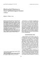
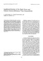
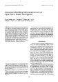
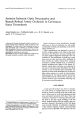
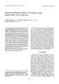
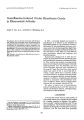
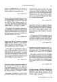
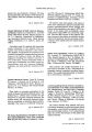
Show Journal of Clinical Neuro- ophthalmology 10( 3): 185- 187, 1990. © 1990 Raven Press, Ltd., New York Aneurysm Mimicking Intracranial Growth of Optic Nerve Sheath Meningioma Klara Landau, M. D., Jonathan c. Horton, M. D., Ph. D., William F. Hoyt, M. D., and Charles B. Wilson, M. D. Summary: A patient with a 3D- year history of blindness in the right eye developed progressive temporal visual loss in the left eye. Examination showed right optic atrophy with optociliary shunts and left band atrophy. These clinical findings suggested that the visual deficit was caused by a right optic nerve sheath meningioma that had grown intracranially to involve the chiasm. Magnetic resonance imaging and surgical exploration revealed a perioptic meningioma extending from the orbit through the optic canal and over the tuberculum sellae. The tumor did not impinge on the optic chiasm or the left optic nerve. The chiasm was compressed by a thrombosed giant right internal carotid artery aneurysm. Key Words: Optic nerve sheath meningioma- Internal carotid artery aneurysm- Chiasmal compression. From the Neuro- Ophthalmology Unit, Departments of Neurological Surgery, Neurology, and Ophthalmology. ( K.~., ]. C. H., W. F. H.), School of Medicine, University of Califorrua, San Francisco, California, and the Neurosurgery Department ( C. B. W.), School of Medicine, University of California, San Francisco, California. Dr. Landau is a recipient of an American Physicians Fellow-ship. . . Address correspondence and reprint requests to Dr. William F. Hoyt, Neuro- Ophthalmology Unit, Room U125, School of Medicine, University of California, San Francisco, CA, 941430350, U. S. A. 185 The association of intracranial aneurysms and meningiomas has been described repeatedly. Occasionally they occur at the same site and both contribute to symptoms ( 1,2). This report describes what we believe is a unique association of a right optic nerve sheath meningioma and a giant right internal carotid artery aneurysm. The meningioma caused blindness in the right eye; progressive temporal field loss in the left eye was due to the aneurysm. CASE REPORT A 59- year- old man reported painless loss of vision in his right eye beginning in 1960. He was seen by several ophthalmologists, but the cause of his progressive visual loss was not determined. He was well until December 1982, when he had a stroke causing left hemiparesis with left lower facial weakness. He also complained of blurred vision in his left eye. Visual field examination performed on a Dicon Auto- Perimeter in January 1983 showed a superior temporal quadrant defect respecting the vertical meridian. As the right eye was blind, it was assumed that the temporal field defect was part of a homonymous field defect caused by the stroke. The patient recovered normal motor function, but a repeat visual field examination of the left eye in 1985 showed the same superior temporal defect. In early 1989 the patient reported progression of temporal field loss in his left eye. He was seen by an ophthalmologist on August 3, 1989. The right eye had no light perception. The left eye saw 20/ 20with + 2.5 sphere correction. A full threshold central 30- 2 Humphrey visual field examination performed using a + 5.0 spherical lens revealed a complete temporal hemianopia and a lens rim artefact in the inferior nasal field ( Fig. 1). The patient 186 K. LANDAU ET AL. FIG. 1. Left visual field showing temporal hemianopia and inferior nasal lens rim artefact. A 30- 2 full thresh~ Id test w~ s performed on a Humphrey automated perimeter wIth a + 5.0 sphere correction. was referred to University of California Medical Center ( San Francisco) for further evaluation and management. When first examined at the Neuro- Ophthalmology Unit on August 24, 1989, the patient had no light perception in his right eye and 20/ 30 acuity in the left eye. There was mild right eye proptosis. The right optic disc was atrophic with several optociliary shunt vessels. The left disc had band atrophy. A tangent screen examination showed a nearly total temporal hemianopia. Optic atrophy with optociliary shunts indicated that the blindness in the right eye was caused by an orbital perioptic meningioma. The progressive temporal visual field defect and band atrophy of the left disc suggested intracranial growth of the perioptic meningioma to involve the chiasm. Magnetic resonance scans confirmed an mtraorbital lesion surrounding the right optic nerve that enhanced with Gadolinium. It extended from the globe to the right optic canal and resembled an optic nerve sheath meningioma. There was a hypointense lesion in the right parieto- occipital lobe representing an old infarct. In addition, a large lobulated mass was present above the right suprasellar cistern with low intensity signal on T1 images and heterogeneous signal intensity on T2 images ( Fig. 2). Angiographically this was a partially thrombosed right carotid aneurysm. The calcified wall of the aneurysm was visible on a lateral skull film ( Fig. 3). Left frontal craniotomy revealed a small meningioma emerging from the intracranial meatus of the right optic canal, but not involving chiasm or left optic nerve. The dilated right internal carotid artery bulged medially beneath the right optic nerve and chiasm. A calcified giant aneurysm originated from the lateral wall of the supraclinoid internal carotid artery and extended laterally. To relieve compression of the chiasm, the right optic nerve was cut a few mm from the optic canal, allowing the chiasm to shift slightly to the left, away from the aneurysm. One week after surgery, visual acuity in the left eye remained 20/ 30 and the tangent screen visual field showed the same dense temporal hemianopia as before. A full threshold central 30- 2 Humphrey visual field showed essentially no change in the patient's visual performance. Another examination 6 weeks after surgery was identical. DISCUSSION Intracranial meningiomas represent 1~ 18% of all primary intracranial tumors ( 3). An intracranial FIG. 2. Left: T1 weighted axial MR ( magnetic resonance) scan showing Gadolinium enhanced right sided optic nerve sheath meningioma ( thin arrow) with small intracranial component ( thick arrow). Right: T2 weighted axial MR scan showing partially thrombosed right suprasellar giant carotid artery aneurysm. ANEURYSM AND OPTIC MENINGIOMA 187 FIG. 3. Left: Lateral skull film of sellar region showing faint dumbbell- shaped calcification ( arrows) on the wall of the giant aneurysm. Right: Lateral selective right internal carotid artery angiogram showing irregular fusiform aneurysm of cavernous and supraclinoid internal carotid artery. aneurysm is found in less than 2% of the population and is symptomatic in less than 1% ( 4). These figures are high enough to lead to an occasional, purely incidental occurrence of both conditions. This association was first described by Arieti in 1944 ( 5). Plangger and co- authors ( 6) recently summarized the literature on 50 such cases, none of which included a primary orbital perioptic meningioma. Most reports describe single cases except for two. Pia et al. ( 1) collected 25 cases from his own experience, by questionnaire sent to his colleagues, and from previous reports. Yasargil described 6 of his own cases ( 7). The latter two studies suggested that the association of meningiomas with aneurysms was more common than could be expected from pure chance alone ( the same was found for pituitary adenomas and aneurysms). Several modes of presentation have been described. Usually the tumor causes the symptoms ( 71% in Pia's series, 83% in Yasargil's series). Less frequently the aneurysm is responsible ( 25% in Pia's series, 17% in Yasargil's series). Occasionally both lesions contribute to symptoms ( 4% in Pia's series, no case in Yasargil's series, a single case report by Love [ 2]). Interestingly, the optic nerve sheath meningioma in our patient began to cause visual loss in the right eye 30 years before a diagnosis was established, and deterioration of vision in the contralateral eye was caused by a second, unrelated lesion. A similar situation was described by Adson in 1941 ( 8). One of his patients ( case 5) was a 42- year- old man who suffered visual loss from a pituitary macroadenoma. It was removed. Tumor recurrence was suggested 7 years later when vision deteriorated. A second craniotomy disclosed no tumor. Instead, a fusiform aneurysm of left internal carotid artery was present, compressing the left optic nerve against the roof and the mesial wall of the optic canal. REFERENCES 1. Pia HW, Obrador S, Martin JG. Association of brain tumours and arterial intracranial aneurysms. Acta Neurochir 1972; 27: 189- 204. 2. Love JG. Association of carotid aneurysm with neoplasm compressing the optic chiasm. Mayo Clin Proc 1963; 38: 15661. 3. Miller NR. Tumors of the meninges and related tissues. In: Walsh and Hoyt's Clinical Neuro- ophthalmology; Vol. 3, 4th ed. Baltimore: Williams & Wilkins, 1988: 1325- 79. 4. Weir B. Intracranial aneurysms and subarachnoidal hemorrhage: an overview. In: Wilkins RH, Rengachary S, eds. Neurosurgery; Vol. 2. New York: McGraw- HilI Book Co, 1985: 1308- 29. 5. Arieti S. Multiple meningioma and meningiomas associated with other brain tumors. I Neuropathol Exp Neural 1944; 3: 255- 70. 6. Plangger CA, Twerdy K. Mohsenipour L Grunert V. Gleichzeitiges Vorkommen von Himtumoren und Aneurysmen. Nervenarzt 1987; 58: 279-- 86. 7. Yasargil GM. Association of brain tumors and cerebral aneurysms. In: Microneurosurgery; Vol. 1. Stuttgart, New York: Georg Thieme Verlag, 1984: 318- 20. 8. Adson AW. Surgical treatment of vascular diseases altering the function of the eyes. Trans Am Acad Ophthalmol OtolaryngoI1941; 46: 95- 111. J Clin Neuro- ophtlullmol, Vol. 10, No. 3, 1990 |