| OCR Text |
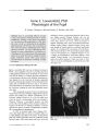
Show VIEWPOINT Pituitary Apoplexy Goes to the Bar: Litigation for Delayed Diagnosis, Deficient Vision, and Death Noble J. David, MD Abstract: Over the past 15 years, I have reviewed the records of six patients whose physicians were alleged to have failed to diagnose a pituitary tumor before it bled (" apoplexy") or failed to recognize apoplexy after it had occurred. These cases were referred by attorneys involved in lawsuits alleging medical negligence. Their histories are summarized here from available clinical records, depositions, and other legal documents. None of these cases is still in litigation. The purpose of this commentary is to show how such legal case material is useful in alerting the medical community to pitfalls in diagnosis. (/ Neuro- Ophthalmol 2006; 26: 128- 133) any physicians are unfamiliar with pituitary apoplexy, the syndrome of acute swelling caused by bleeding or infarction within a pre- existing macroadenoma. Severe headache with a variable mix of acute visual loss, ocular motor dysfunction, and meningeal signs typify this emergency. Diagnostic confusion and delay in its treatment may lead to permanent visual loss or death ( see the Warwar article on page 95 in this issue of the journal) ( 1- 3). The modern era of pituitary oncology began in the 1880s with reports ( 4) associating acromegaly with pituitary neoplasm. Bailey ( 5) in 1897 and Bleibtreu ( 6) in 1904 described acromegalic patients whose death was caused by hemorrhage into a pituitary tumor. In 1950, Brougham, Heusner, and Adams ( 7) published their seminal clinico-pathologic report of five patients who died of pituitary hemorrhage or infarction and dubbed this complication " pituitary apoplexy." Although their clinical vignettes epitomized quite accurately the signs and symptoms of this emergency, it was not until the mid 1970s that reports addressed the spectrum of clinical manifestations ( 1- 3,8) and the crucial role of the newly introduced CT in Department of Neurology, University of Miami, Miami, Florida. Address correspondence to Noble J. David, MD, 1519 Granada Boulevard, Coral Gables, FL 33134; E- mail: nobbyd@ earthlink. net corroborating this diagnosis ( 9,10). Verrees et al ( 11) have recently published a comprehensive clinical summary and thorough bibliography on this subject. CASE 1: NO INSURANCE, NO CARE A 48- year- old woman consulted her physician for headache and vomiting of 24 hours' duration. He diagnosed " migraine" but requested a brain CT scan, which her health plan refused to authorize. She was evaluated the next day at a hospital emergency room for increasing headache, vomiting, and photophobia but left prematurely when she was told that her insurance would not pay for treatment. That evening and again on the third day of illness, she visited a " drop- in" clinic, where her persistent complaints were diagnosed as " migraine with urinary tract infection." She was described as " oriented, neck supple but drowsy from her medication." She was given an appointment for neurologic consultation and CT to be done three days later. On the fourth day of illness, her sister escorted her to yet another emergency room, where, in addition to her unabated head pain and emesis, she reported " blurred and double vision." She was described by the nurse as arousable but drowsy with a fever of 101.5° F. Examination performed by a general physician three hours later showed that both pupils were 3 mm and unreactive to light. The physician recorded " lateral strabismus" and could demonstrate vision no better than hand movements in either eye. Emergency brain CT scan was interpreted as showing " a 2.8 cm mass of homogenous density in the suprasellar area ... probable aneurysm, without evidence of subarachnoid hemorrhage." The physician telephoned a neurosurgical colleague who diagnosed pituitary apoplexy from the clinical description and recommended emergency trans- sphenoidal surgery. He could not accept her as a patient because her health plan did not cover his services. She was started on methylprednisolone intravenously and transferred to another hospital, where MRI revealed a hemorrhagic pituitary adenoma extending into the suprasellar space and compressing the optic chiasm. She underwent trans- sphenoidal evacuation of the tumor, but 128 J Neuro- Ophthalmol, Vol. 26, No. 2, 2006 Pituitary Apoplexy Litigation J Neuro- Ophthalmol, Vol. 26, No. 2, 2006 the left eye visual acuity thereafter was no light perception and the right eye visual acuity was 20/ 60 with a constricted nasal field. Legal Aspects Most of the physicians and clinics involved in her care before operation were sued for delayed diagnosis. Her emergency treatment was critically compromised by refusal of the health care organizations, which were legally immune, to authorize the initial CT or treatment outside her plan. The malpractice claims were settled for several million dollars. Medical Aspects Pituitary apoplexy was not suspected at a succession of clinics and emergency rooms. That the correct diagnosis was made by a telephone consultation illustrates the importance of familiarity with this condition. CASE 2: DEATH IN THE DOCTOR'S OFFICE A 43- year- old construction worker was treated by his family physician for headaches ascribed to hypertension. No visual complaints were recorded. Six months later, he awoke febrile, perspiring, and complaining of headache and extreme generalized weakness. His family took him to the doctor's office, where he lay in the treatment room for three hours. His wife then noted rapid and laborious breathing. Within 20 minutes, he became unresponsive and apneic. He could not be resuscitated. At autopsy, the medical examiner found suprasellar extension of a large chromophobe adenoma compressing the optic chiasm and hypothalamus. The tumor was edematous and peppered with petechial hemorrhages. Legal Aspects The widow lodged a complaint against the physician for not suspecting a neurologic cause for the patient's headache and failure to act promptly in his final illness. Defense attorneys for the physician found records revealing that the patient had been studied three years earlier in another city by an ophthalmologist who recorded visual field defects classic for compression of the optic chiasm. CT scan at that time had revealed a pituitary tumor with suprasellar extension. When told that he must undergo surgical and/ or radiation therapy, he had demurred. The attending physician had not obtained his history and blamed himself for not diagnosing the condition. The case was settled out of court. Medical Aspects This history cautions the practitioner that large suprasellar tumors may produce no obvious endocrine or neurologic symptoms. Patients known to harbor such lesions must be warned that abrupt headache or visual loss should cause them to seek emergency neurosurgical attention. CASE 3: MISDIAGNOSIS OF GLAUCOMA AND MIGRAINE A 41 - year- old laborer visited an emergency room for headache of several weeks' duration, which had become intense. He was told he had migraine, was given an opiate injection, and sent home. The next morning his wife called the fire rescue service when she could not get him out of bed, noting that he was weak on his left side. He was admitted to a hospital, where a neurologist found him to be febrile and lethargic but responsive to simple commands. His neck was stiff and his left extremities weak and hypotonic. His pupils were 5 mm and unreactive to light. Both optic fundi were described as normal. Eye movements were said to be full to passive head rotation. A brain CT without contrast was reported as showing " an ovoid area of decreased absorption within the inferior right frontal lobe with minimal mass effect on the anterior horn." Lumbar puncture revealed an opening pressure of 490 mm water and yielded turbid xanthochromic fluid with 200 polymorphonuclear cells/ cm3. The cerebral arteriogram was read as showing " spasticity and elevation of the anterior cerebral and supraclinoid carotid arteries. Anterior communicating aneurysm a possibility." His manifestations were attributed to subarachnoid hemorrhage with vasospasm and cerebral infarction. However, because of the possibility of bacterial meningitis, he was given intravenous antibiotics. Five hours after his admission to the hospital, spontaneous respirations ceased. He was declared brain dead, and life support was terminated. Autopsy revealed a large hemorrhagic suprasellar chromophobe adenoma stretching the optic chiasm and compressing the hypothalamus. Subarachnoid blood was found in the middle fossa and cisterns. The entire brain was edematous. Legal Aspects After the patient's death, a plaintiff attorney obtained the following information: for the five months preceding his death, the patient had complained to his primary care physician of headaches and visual blurring. He had been sent to an ophthalmologist who, on three visits, had found abnormal visual fields and interpreted them as consistent with glaucoma. Three days before his death, paranasal sinus films ordered by an otolaryngologist were read as normal but were later interpreted as showing sellar erosion and expansion. All physicians involved in the patient's care 129 J Neuro- Ophthalmol, Vol. 26, No. 2, 2006 David were sued, but the ophthalmologist was held most liable for missing the diagnosis. Each physician settled out of court. Medical Aspects Pituitary apoplexy is commonly misdiagnosed as migraine or subarachnoid hemorrhage but infrequently mimics meningeal infection. Hemiparesis is rare in pituitary apoplexy but may occur because compression of the internal carotid or middle cerebral artery results from swelling of a tumor encasing these vessels ( 12). CASE 4: FALSE- NEGATIVE CT FINDING A 55- year- old school teacher consulted her doctor for recent- onset headaches. Three months earlier, an ophthalmologist had found elevated intraocular pressure in both eyes, for which he had performed bilateral laser iridectomies. At that time she had no visual symptoms, and visual acuity was 20/ 20 in both eyes. Visual fields were not recorded. Nocturnal headaches increased in severity, unrelieved by analgesics. Her doctor ordered a brain CT that was reported by the radiologist as normal. Seven months later, she had abrupt development of a severe holocranial headache, the worst in her life. She sought help in a hospital emergency room, where she was thought to have migraine, was given intravenous medication, and sent home. A few hours later, her daughter took her to an ophthalmologist because her vision was failing. He found " hand movements only in one nasal field." On admission, the initial diagnosis was migraine, meningitis, or posterior hemisphere infarction. Brain CT revealed a large suprasellar extension of a pituitary adenoma in which MRI showed multiple hemorrhagic foci. On high doses of intravenous dexamethasone, the patient's vision improved slightly over the next five days. She was then referred to a neurosurgeon, who performed a transfrontal craniotomy. Her postoperative course was complicated by cerebrospinal rhinorrhea, requiring a second craniotomy. She was left with a dense bitemporal hemi-anopia and visual acuities of 20/ 100 right eye and 20/ 20 left eye. Legal Aspects She sued the radiologist after independent review of her first CT scan identified an intrasellar mass typical of pituitary adenoma with suprasellar extension. ( The emergency room physician who sent her out with the diagnosis of migraine was not sued.) An out- of- court settlement was reached. Medical Aspects The primary symptom was chronic headache, doubtless related to the suprasellar tumor, given that she had had no prior headache history and that the headache disappeared after surgery. The apoplectic presentation provoked common diagnostic missteps. CT and MRI eventually revealed the diagnosis, which had not been suspected clinically. CASE 5: IMPOTENCE FAILS TO TRIGGER AN INTRACRANIAL EVALUATION A 49- year- old store manager consulted a urologist for inability to achieve an erection. He was diagnosed as having Peyronie disease and phimosis. He had had a vasectomy eight years before. Testing revealed a low testosterone level but normal T4, FSH, LH, and prolactin levels. Testosterone injections restored his potency, but he complained of severe bitemporal headaches during coitus. No follow- up examinations occurred until nine months later, when he was awakened by severe bitemporal headaches followed by vomiting and double vision. On emergency room examination, he was described as pale, bradycardic, and obtunded. He had nuchal rigidity but was afebrile. Visual acuity was normal in both eyes, but a right lateral rectus palsy was noted. Brain non- contrast CT was interpreted as normal. Lumbar puncture revealed colorless, acellular fluid; opening pressure was not recorded. The normal spinal fluid confounded his doctors, who believed that he had had a subarachnoid hemorrhage. The next day, he had a right ptosis and third cranial nerve palsy, which prompted a change in his diagnosis to " occlusive vertebrobasilar disease." He was treated with intravenous dexamethasone and heparin. Repeat brain CT scan with contrast showed a 2.8 cm suprasellar mass, which in retrospect, was present in the first CT scan. A pituitary tumor was diagnosed ( there was no mention of apoplexy). Five days after admission, the patient underwent right frontal craniotomy for removal of the tumor, which, on histologic study, was a chromophobe adenoma with multiple foci of necrosis. After the operation, he received 5,000 rads of gamma radiation to the sellar area. His ocular motor defects cleared steadily and completely. He did well on replacement thyroid, corticosteroid, and androgen but had permanent, total loss of his sense of smell. Legal Aspects Because of persistent anosmia, the patient sued the urologist for not investigating his pituitary status in the course of studying his impotence. He argued that earlier diagnosis might have allowed a trans- sphenoidal rather than a transcranial approach and would thereby have preserved his sense of smell. A defense expert argued that pituitary tumors are rarely identified through the solitary complaint of impotence and that there was no other damage. The jury found in favor of the defendant. 130 © 2006 Lippincott Williams & Wilkins Pituitary Apoplexy Litigation J Neuro- Ophthalmol, Vol. 26, No. 2, 2006 Medical Aspects Asthenia, impotence, amenorrhea, and loss of sexual habitus are often interpreted by the attending physician as primary hypothyroidism or gonadal failure rather than hypopituitarism. CASE 6: APOPLEXY ON THE EVE OF NEUROSURGICAL APPOINTMENT A 41- year- old construction worker visited his optometrist for a " spot of blurred vision" in his right eye. Nine months earlier, normal visual acuity and visual fields had been recorded in that office. He was referred to a retinal specialist to whom he reported " visual distortion." The specialist found normal visual acuity in both eyes, but on Amsler grid testing, the patient described temporal blurring between fixation and the blind spot in the right eye. Ophthalmoscopy and fluorescein fundus angiography were normal. Even so, he was given a diagnosis of central serous chorioretinopathy, perhaps because his visual complaint sounded to the specialist like metamorphopsia. Six weeks later, the patient's vision markedly worsened. Visual fields showed bitemporal hemianopia. He was referred to a neuro- ophthalmologist who ordered an MRI that revealed a large intrasellar mass with suprasellar extension, diagnosed as a pituitary macroadenoma. Serum prolactin was normal. He was scheduled to be seen by a neurosurgeon one week later. On the eve of this appointment, he was awakened from sleep by the worst headache of his life. In the emergency room of a nearby hospital, he was examined by another neurosurgeon, who ordered a brain CT scan that confirmed the presence of the suprasellar tumor. The patient was given narcotic analgesics and sent out with instructions to keep his appointment with the neurosurgeon whom he had been scheduled to see. No mention was made of the possibility of pituitary apoplexy. By morning he was vomiting, febrile, and confused. The headache was unrelieved. His wife brought him to the neurosurgeon to whom he had initially been referred. He was blind except for a left upper nasal quadrant of vision and had a " bilateral incomplete third cranial nerve deficit." The next day, he was admitted with a diagnosis of probable berry aneurysm. After a two- day delay, during which cerebral angiography failed to confirm the presence of a brain aneurysm, he underwent transcranial surgery for removal of a necrotic and hemorrhagic pituitary adenoma. The postoperative course was stormy with subdural, epidural, and subgaleal hematomas requiring two additional craniotomies and insertion of a ventriculostomy, all within one week of the initial surgery. The patient gradually recovered but was left with only a nasal quadrant of vision in the left eye. Legal Aspects The patient sued the retinal specialist for delay in diagnosis and sued the neurosurgeon for sending him out of the emergency room. The neuro- ophthalmologist was also named in the complaint, although he had made the appropriate diagnosis and referral. All suits were settled out of court for undisclosed amounts. Medical Aspects Judging from the swift progression of his visual symptoms, the patient's tumor was rapidly enlarging. The patient's initial complaint of distorted vision, a hallmark of retinal disease, may have been misleading. Ironically, apoplexy struck on the eve of his neurosurgical appointment. Transcranial surgery was delayed two days to rule out berry aneurysm. DISCUSSION Claims of medical negligence arose not only from allegations of mismanagement of the apoplectic episode but from failure to diagnose the tumor before apoplexy. Pituitary tumors are discovered in one of five ways: 1. Hyperpituitarism: The dramatic endocrine caricatures of acromegaly and Cushing disease reflect hypersecretion of growth hormone or ACTH, respectively, and provide direct clinical evidence of a hypersecretory adenoma. Prolactinomas, which cause the infertility- galactorrhea-amenorrhea syndrome, can be diagnosed by assaying serum prolactin. They are unique in that tumor growth and secretion can be controlled medically with bromocriptine or its analogs. 2. Hypopituitarism: Hyposecretion of one or more of the hormones caused by compression of the functioning gland by a non- secreting tumor. Patients with mild hypothyroidism or hypogonadism are often treated with thyroid or sex hormones without being evaluated for pituitary dysfunction ( Case 5). 3. Progressive visual loss: Chiasmal compression occurs only after the tumor has grown more than 1 cm above the diaphragma sellae. Such patients are often given new glasses or the symptoms are ascribed to cataract or glaucoma. Patients with amblyopia in one or both eyes not well explained by ophthalmologic findings ( Cases 2 and 6) deserve study with visual fields followed by brain imaging. Failure to detect bitemporal hemianopia, which is pathognomic for chiasmal damage, is regularly cited as negligence. 4. CT and MRI: Most pituitary adenomas are now discovered fortuitously by these imaging studies in screening patients referred on suspicion of other intracranial disease. The unanticipated lesion, which has been called an " incidentaloma"( 13), is usually unrelated to 131 J Neuro- Ophthalmol, Vol. 26, No. 2, 2006 David the symptoms being studied. Because of the threat of apoplexy, it is important to treat even asymptomatic adenomas promptly ( see Case 3, who proceeded to apoplexy after the tumor was overlooked on an earlier CT scan). Chronic headaches in patients ( Cases 2, 3, and 4) with rapidly enlarging suprasellar adenomas are best explained by stretching of the pain- sensitive sellar diaphragm. Nonetheless, many patients with large pituitary tumors, including those with visual involvement, do not complain of headache, probably because enlargement occurs gradually. 5. Pituitary apoplexy: Apoplexy occurs in approximately 10% of untreated patients, most of whom are unaware that they have the tumor. Precipitating factors such as anticoagulation, head trauma, or surgery under general anesthesia have been implicated, but most patients lack such a history. The clinical syndrome of apoplexy is the result of abrupt expansion caused by bleeding and/ or infarction in the tumor. The cardinal symptom is holocranial headache of sufficient severity to suggest bleeding aneurysm or acute meningitis, usually described as " the worst of my life," and often worse on the side of visual or ocular motor involvement. Neuro-ophthalmologic findings are paramount, including a combination of chiasmal visual loss and palsies of the extraocular muscles, the latter rarely seen in chronically enlarging adenomas without hemorrhage. In contrast to the isolated unilateral third cranial nerve palsy typical of posterior communicating aneurysm, the patient with pituitary apoplexy may display any combination of third, fourth, or sixth cranial nerve involvement on either or both sides, findings that may also simulate basilar infarction. The headache may be falsely attributed to subarachnoid hemorrhage from berry aneurysm or migraine as in Cases 1, 3, and 4. In pituitary apoplexy, symptoms of meningeal irritation result from leakage of blood or necrotic debris into the subarachnoid space. Lumbar puncture reveals elevated pressure and yields either bloody and xanthochromic fluid or a pleocytosis of variable intensity and cell type, suggesting a bacterial or viral meningitis. The most serious complications of pituitary apoplexy are blindness and hyperpyrexic coma from suprasellar expansion compressing the optic pathways and the hypothalamus. Visual fields by finger confrontation may demonstrate relatively profound temporal field loss, even in patients who have severe visual impairment. Optic disc pallor is evidence of chronic optic nerve or chiasmal damage before apoplexy. The diagnosis is clinched by demonstration on a non-contrast brain CT of a large adenoma that may or may not show intrinsic changes of bleeding or necrosis. The scan is often of poor technical quality because the patient's pain and obtundation limit cooperation. MRI offers superior resolution of the pathologic anatomy of pituitary tumors, including apoplectic changes, but is rarely available for immediate use in the emergency room. Consultative review of all the findings with other specialists, including the neurologist, ophthalmologist, and endocrinologist, may bring help from doctors familiar with this disorder. Intravenous corticosteroids in high dosage ( methylprednisolone 500 mg q4h) must be given promptly and may effect dramatic clinical relief. Antibiotics are indicated in patients with suspected bacterial meningitis until the diagnosis is settled. The patient must be monitored continuously for abrupt clinical change. As in subarachnoid hemorrhage, patients with pituitary apoplexy require immediate referral to the neurosurgeon. If vision is threatened, the tumor should be decompressed without delay, preferably by the trans- sphenoidal route. Of the three patients who underwent transfrontal craniotomy ( Cases 1, 4, and 6), two had stormy postoperative courses requiring re- operation. All were left with anosmia. Unlike the defects in ocular motility, which usually recover completely, visual loss from chiasmal compression of more than 24 hours is usually irreversible. CONCLUSIONS In 1912, the legendary neurosurgeon Harvey Cushing wrote in the preface to his remarkably prescient book " The Pituitary Body and its Disorders" ( 14) that disease in this gland concerns a wide assortment of specialists, an observation that remains valid almost 100 years later. Because pituitary apoplexy is a treatable entity that threatens sight and survival, plaintiff attorneys pursuing claims of medical negligence often secure large sums in settlements or awards in these lawsuits. Few physicians regard the deposition or courtroom experience as an appropriate venue for continuing medical education, but I submit that scrutiny of the legal documents, as well as the clinical records, may warn us of common and predictable pitfalls. The curriculum of continuing medical education could be further enhanced by presentations that share with the medical community the experiences of physicians who have served as expert witnesses in malpractice lawsuits involving high- risk diagnoses. Their observations would help to improve patient treatment and to forestall legal challenges. REFERENCES 1. David NJ, Gargano FP, Glaser JS. Pituitary Apoplexy in Clinical Perspective. In: Glaser JS, Smith JL, eds. Neuro- Ophthalmology, vol VIII. St. Louis: Mosby; 1975: 140- 165. 2. Post MJ, David NJ, Glaser JS, et al. Pituitary apoplexy: diagnosis by computed tomography. Radiology 1980; 134: 665- 70. 132 © 2006 Lippincott Williams & Wilkins Pituitary Apoplexy Litigation J Neuro- Ophthalmol, Vol. 26, No. 2, 2006 3. Ostrov SG, Quencer RM, Hoffman JC, et al. Hemorrhage within pituitary adenomas: how often associated with pituitary apoplexy syndrome? AM Am J Roentgenol 1989; 153: 503- 10. 4. Cushing H. The Pituitary Body and Its Disorders. Birmingham: Classics of Medicine Library; 1979: 27. 5. Bailey P. Pathological report of a case of akromegaly, with special reference to lesions in the hypophysis cerebri, etc. The Philadelphia Medical Journal 1897; 1: 789- 92. 6. Bleibtreu, L. Ein Fall von akromegalie ( Zerstorung der hypophysis durch blutung). Muenchener Medizinische Wochenschrift 1904; 43: 279- 80. 7. Brougham M, Heusner AP Adams RD. Acute degenerative changes in adenomas of the pituitary body with special reference to pituitary apoplexy. J Neurosurg 1950; 7: 421- 39. 8. Rovit RL, Fein JM. Pituitary apoplexy: a review and reappraisal. J Neurosurg 1972; 37: 280- 8. 9. Conomy JP, Ferguson JH, Brodkey JS, et al. Spontaneous infarction in pituitary tumors: neurologic and therapeutic aspects. Neurology 1975; 25: 580- 7. 10. Weisberg LA. Pituitary apoplexy: association of degenerative change in pituitary adenoma with radiotherapy and detection by cerebral computed tomography. Am J Med 1977; 63: 109- 15. 11. Verrees M, Arafa BM, Selman WR, et al. Pituitary tumor apoplexy: characteristics, treatment and outcomes. Neurosurg Focus 2004; 16: E6. 12. Schnitker MT, Lehnert HB. Apoplexy in a pituitary chromophobe adenoma producing the syndrome of middle cerebral artery thrombosis: case report. J Neurosurg 1952; 9: 210- 3. 13. Molitch ME, Russel EJ. The pituitary ' incidentaloma'. Ann Intern Med 1990; 112: 925- 31. 14. Cushing H. The Pituitary Body and Its Disorders. Birmingham: Classics of Medicine Library; 1979: Preface, ix. 133 |